Movie
Movie Controller


+ Open data

- Basic information
| Entry |  | |||||||||
|---|---|---|---|---|---|---|---|---|---|---|
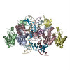
| Title | HIV-1 wild-type intasome core | |||||||||
 Map data Map data | Primary map | |||||||||
 Sample Sample |
| |||||||||
 Keywords Keywords | HIV-1 / integrase / nucleoprotein complexes / INSTI / VIRAL PROTEIN | |||||||||
| Function / homology |  Function and homology information Function and homology informationDNA integration / viral genome integration into host DNA / establishment of integrated proviral latency / RNA stem-loop binding / RNA-directed DNA polymerase activity / endonuclease activity / DNA recombination / symbiont entry into host cell / DNA binding / zinc ion binding Similarity search - Function | |||||||||
| Biological species |   Human immunodeficiency virus 1 Human immunodeficiency virus 1 | |||||||||
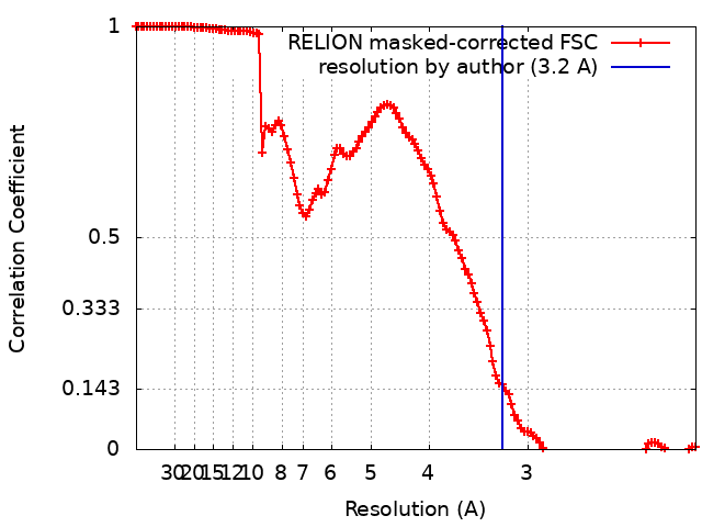
| Method | single particle reconstruction / cryo EM / Resolution: 3.2 Å | |||||||||
 Authors Authors | Li M / Craigie R | |||||||||
| Funding support |  United States, 1 items United States, 1 items
| |||||||||
 Citation Citation | Journal: J Mol Biol / Year: 2024 Title: HIV-1 Integrase Assembles Multiple Species of Stable Synaptic Complex Intasomes That Are Active for Concerted DNA Integration In vitro. Authors: Min Li / Renbin Yang / Xuemin Chen / Huaibin Wang / Rodolfo Ghirlando / Emilios K Dimitriadis / Robert Craigie / Abstract: Retroviral DNA integration is mediated by nucleoprotein complexes (intasomes) in which a pair of viral DNA ends are bridged by a multimer of integrase (IN). Most of the high-resolution structures of ...Retroviral DNA integration is mediated by nucleoprotein complexes (intasomes) in which a pair of viral DNA ends are bridged by a multimer of integrase (IN). Most of the high-resolution structures of HIV-1 intasomes are based on an HIV-1 IN with an Sso7d protein domain fused to the N-terminus. Sso7d-IN aggregates much less than wild-type IN and has been critical for structural studies of HIV-1 intasomes. Unexpectedly, these structures revealed that the common core architecture that mediates catalysis could be assembled in various ways, giving rise to both tetrameric and dodecameric intasomes, together with other less well-characterized species. This differs from related retroviruses that assemble unique multimeric intasomes, although the number of protomers in the intasome varies between viruses. The question of whether the additional Sso7d domain contributes to the heterogeneity of HIV-1 intasomes is therefore raised. We have addressed this by biochemical and structural studies of intasomes assembled with wild-type HIV-1 IN. Negative stain and cryo-EM reveal a similar range of multimeric intasome species as with Sso7d-IN with the same common core architecture. Stacks of intasomes resulting from domain swapping are also seen with both wild-type and Sso7d-IN intasomes. The propensity to assemble multimeric intasome species is, therefore, an intrinsic property of HIV-1 IN and is not conferred by the presence of the Sso7d domain. The recently solved intasome structures of different retroviral species, which have been reported to be tetrameric, octameric, dodecameric, and hexadecameric, highlight how a common intasome core architecture can be assembled in different ways for catalysis. | |||||||||
| History |
|
- Structure visualization
| Supplemental images |
|---|
- Downloads & links
-EMDB archive
| Map data | emd_43705.map.gz | 158.7 MB | EMDB map data format | |
|---|---|---|---|---|
| Header (meta data) | emd-43705-v30.xmlemd-43705.xml | 21.5 KB 21.5 KB | Display Display | EMDB header |
| FSC (resolution estimation) | emd_43705_fsc.xml | 12.7 KB | Display | FSC data file |
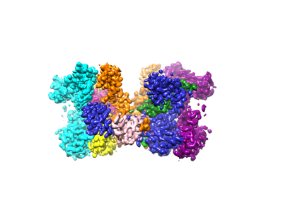
| Images |  emd_43705.png emd_43705.png | 65.1 KB | ||
| Filedesc metadata | emd-43705.cif.gz | 6.5 KB | ||
| Others | emd_43705_additional_1.map.gzemd_43705_half_map_1.map.gzemd_43705_half_map_2.map.gz | 159.1 MB 159.5 MB 159.5 MB | ||
| Archive directory |  http://ftp.pdbj.org/pub/emdb/structures/EMD-43705ftp://ftp.pdbj.org/pub/emdb/structures/EMD-43705 http://ftp.pdbj.org/pub/emdb/structures/EMD-43705ftp://ftp.pdbj.org/pub/emdb/structures/EMD-43705 | HTTPS FTP |
-Validation report
| Summary document | emd_43705_validation.pdf.gz | 851.9 KB | Display | EMDB validaton report |
|---|---|---|---|---|
| Full document | emd_43705_full_validation.pdf.gz | 851.4 KB | Display | |
| Data in XML | emd_43705_validation.xml.gz | 20.2 KB | Display | |
| Data in CIF | emd_43705_validation.cif.gz | 26.6 KB | Display | |
| Arichive directory | https://ftp.pdbj.org/pub/emdb/validation_reports/EMD-43705ftp://ftp.pdbj.org/pub/emdb/validation_reports/EMD-43705 | HTTPS FTP |
-Related structure data
| Related structure data |  8w09MC 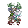 8w2rC 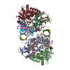 8w34C M: atomic model generated by this map C: citing same article ( |
|---|---|
| Similar structure data |


-Links
| EMDB pages | EMDB (EBI/PDBe) / EMDataResource |
|---|
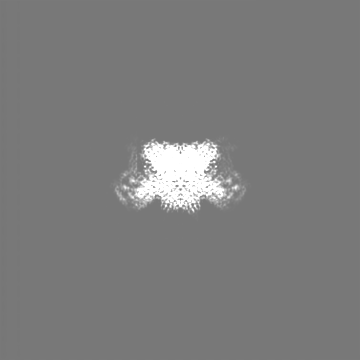
-Map
| File | Download / File: emd_43705.map.gz / Format: CCP4 / Size: 178 MB / Type: IMAGE STORED AS FLOATING POINT NUMBER (4 BYTES) | ||||||||||||||||||||||||||||||||||||
|---|---|---|---|---|---|---|---|---|---|---|---|---|---|---|---|---|---|---|---|---|---|---|---|---|---|---|---|---|---|---|---|---|---|---|---|---|---|
| Annotation | Primary map | ||||||||||||||||||||||||||||||||||||
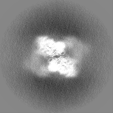
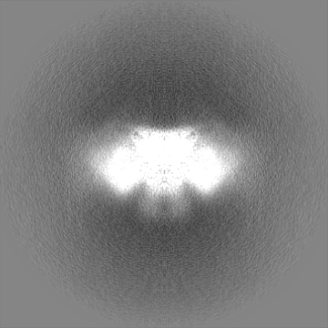
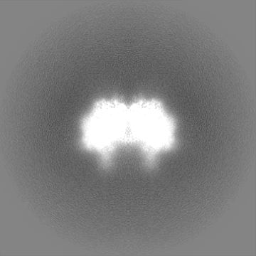
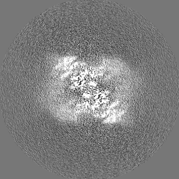
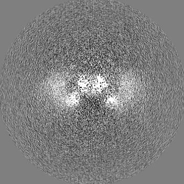
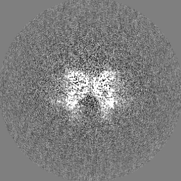
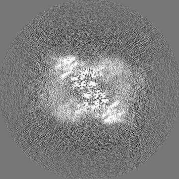
| Projections & slices | Image control
Images are generated by Spider. | ||||||||||||||||||||||||||||||||||||
| Voxel size | X=Y=Z: 1.05 Å | ||||||||||||||||||||||||||||||||||||
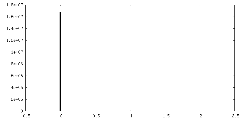
| Density |
| ||||||||||||||||||||||||||||||||||||
| Symmetry | Space group: 1 | ||||||||||||||||||||||||||||||||||||
| Details | EMDB XML:
|
 Z (Sec.)
 Y (Row.)
 X (Col.)


-Supplemental data
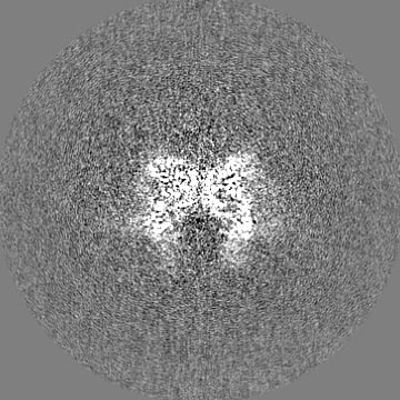
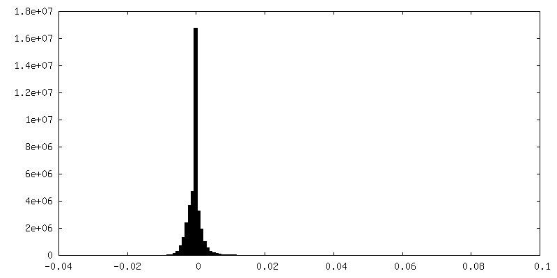
-Additional map: #1
| File | emd_43705_additional_1.map | ||||||||||||
|---|---|---|---|---|---|---|---|---|---|---|---|---|---|
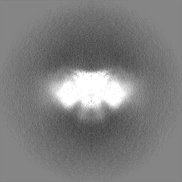
| Projections & Slices |
| ||||||||||||
| Density Histograms |


-Half map: Half1 map
| File | emd_43705_half_map_1.map | ||||||||||||
|---|---|---|---|---|---|---|---|---|---|---|---|---|---|
| Annotation | Half1 map | ||||||||||||
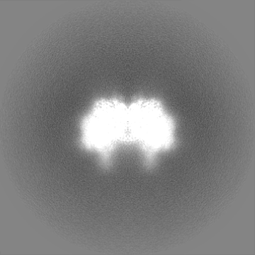
| Projections & Slices |
| ||||||||||||
| Density Histograms |


-Half map: Half2 map
| File | emd_43705_half_map_2.map | ||||||||||||
|---|---|---|---|---|---|---|---|---|---|---|---|---|---|
| Annotation | Half2 map | ||||||||||||
| Projections & Slices |
| ||||||||||||
| Density Histograms |


- Sample components
-Entire : wild-type HIV-1 intasomes
| Entire | Name: wild-type HIV-1 intasomes |
|---|---|
| Components |
|
-Supramolecule #1: wild-type HIV-1 intasomes
| Supramolecule | Name: wild-type HIV-1 intasomes / type: complex / ID: 1 / Parent: 0 / Macromolecule list: #1-#3 Details: HIV-1 intasomes assembled with wild-type NL4-3 integrase |
|---|---|
| Source (natural) | Organism: Human immunodeficiency virus 1 |
| Molecular weight | Theoretical: 400 KDa |
-Macromolecule #1: Integrase
| Macromolecule | Name: Integrase / type: protein_or_peptide / ID: 1 / Number of copies: 8 / Enantiomer: LEVO |
|---|---|
| Source (natural) | Organism: Human immunodeficiency virus 1 |
| Molecular weight | Theoretical: 32.658252 KDa |
| Recombinant expression | Organism:  |
| Sequence | String: GSHMFLDGID KAQEEHEKYH SNWRAMASDF NLPPVVAKEI VASCDKCQLK GEAMHGQVDC SPGIWQLDCT HLEGKVILVA VHVASGYIE AEVIPAETGQ ETAYFLLKLA GRWPVKTVHT DNGSNFTSTT VKAACWWAGI KQEFGIPYNP QSQGVIESMN K ELKKIIGQ ...String: GSHMFLDGID KAQEEHEKYH SNWRAMASDF NLPPVVAKEI VASCDKCQLK GEAMHGQVDC SPGIWQLDCT HLEGKVILVA VHVASGYIE AEVIPAETGQ ETAYFLLKLA GRWPVKTVHT DNGSNFTSTT VKAACWWAGI KQEFGIPYNP QSQGVIESMN K ELKKIIGQ VRDQAEHLKT AVQMAVFIHN FKRKGGIGGY SAGERIVDII ATDIQTKELQ KQITKIQNFR VYYRDSRDPV WK GPAKLLW KGEGAVVIQD NSDIKVVPRR KAKIIRDYGK QMAGDDCVAS RQDED UniProtKB: Gag-Pol polyprotein |
-Macromolecule #2: vDNA
| Macromolecule | Name: vDNA / type: dna / ID: 2 / Number of copies: 2 / Classification: DNA |
|---|---|
| Source (natural) | Organism: Human immunodeficiency virus 1 |
| Molecular weight | Theoretical: 27.371412 KDa |
| Sequence | String: (DA)(DC)(DT)(DG)(DC)(DT)(DA)(DG)(DA)(DG) (DA)(DT)(DT)(DT)(DT)(DC)(DC)(DA)(DC)(DA) (DC)(DT)(DT)(DT)(DT)(DT)(DT)(DT)(DT) (DT)(DT)(DT)(DT)(DT)(DT)(DT)(DT)(DT)(DT) (DT) (DT)(DT)(DT)(DT)(DT)(DT) ...String: (DA)(DC)(DT)(DG)(DC)(DT)(DA)(DG)(DA)(DG) (DA)(DT)(DT)(DT)(DT)(DC)(DC)(DA)(DC)(DA) (DC)(DT)(DT)(DT)(DT)(DT)(DT)(DT)(DT) (DT)(DT)(DT)(DT)(DT)(DT)(DT)(DT)(DT)(DT) (DT) (DT)(DT)(DT)(DT)(DT)(DT)(DT)(DT) (DT)(DT)(DT)(DT)(DT)(DT)(DT)(DT)(DT)(DT) (DT)(DT) (DT)(DT)(DT)(DT)(DT)(DT)(DT) (DT)(DT)(DT)(DT)(DT)(DT)(DT)(DT)(DT)(DT) (DT)(DT)(DT) (DT)(DT)(DT)(DT)(DT)(DT) (DT)(DT)(DT)(DT) |
-Macromolecule #3: vDNA
| Macromolecule | Name: vDNA / type: dna / ID: 3 / Number of copies: 2 / Classification: DNA |
|---|---|
| Source (natural) | Organism: Human immunodeficiency virus 1 |
| Molecular weight | Theoretical: 8.374477 KDa |
| Sequence | String: (DA)(DA)(DA)(DA)(DA)(DA)(DA)(DA)(DG)(DT) (DG)(DT)(DG)(DG)(DA)(DA)(DA)(DA)(DT)(DC) (DT)(DC)(DT)(DA)(DG)(DC)(DA) |
-Macromolecule #4: MAGNESIUM ION
| Macromolecule | Name: MAGNESIUM ION / type: ligand / ID: 4 / Number of copies: 4 / Formula: MG |
|---|---|
| Molecular weight | Theoretical: 24.305 Da |
-Macromolecule #5: ZINC ION
| Macromolecule | Name: ZINC ION / type: ligand / ID: 5 / Number of copies: 2 / Formula: ZN |
|---|---|
| Molecular weight | Theoretical: 65.409 Da |
-Macromolecule #6: (4R,12aS)-N-(2,4-difluorobenzyl)-7-hydroxy-4-methyl-6,8-dioxo-3,4...
| Macromolecule | Name: (4R,12aS)-N-(2,4-difluorobenzyl)-7-hydroxy-4-methyl-6,8-dioxo-3,4,6,8,12,12a-hexahydro-2H-pyrido[1',2':4,5]pyrazino[2,1-b][1,3]oxazine-9-carboxamide type: ligand / ID: 6 / Number of copies: 2 / Formula: DLU |
|---|---|
| Molecular weight | Theoretical: 419.379 Da |
| Chemical component information | 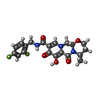 ChemComp-DLU: |
-Experimental details
-Structure determination
| Method | cryo EM |
|---|---|
 Processing Processing | single particle reconstruction |
| Aggregation state | particle |
-Sample preparation
| Concentration | 0.4 mg/mL |
|---|---|
| Buffer | pH: 6.2 |
| Grid | Model: Quantifoil R1.2/1.3 |
| Vitrification | Cryogen name: ETHANE |
- Electron microscopy
| Microscope | FEI TITAN KRIOS |
|---|---|
| Specialist optics | Energy filter - Name: GIF Bioquantum / Energy filter - Slit width: 20 eV |
| Image recording | Film or detector model: GATAN K2 SUMMIT (4k x 4k) / Detector mode: SUPER-RESOLUTION / Average electron dose: 50.0 e/Å2 |
| Electron beam | Acceleration voltage: 300 kV / Electron source:  FIELD EMISSION GUN FIELD EMISSION GUN |
| Electron optics | C2 aperture diameter: 70.0 µm / Illumination mode: FLOOD BEAM / Imaging mode: BRIGHT FIELD / Cs: 2.7 mm / Nominal defocus max: 2.6 µm / Nominal defocus min: 1.4000000000000001 µm / Nominal magnification: 105000 |
| Sample stage | Specimen holder model: FEI TITAN KRIOS AUTOGRID HOLDER / Cooling holder cryogen: NITROGEN |
| Experimental equipment |  Model: Titan Krios / Image courtesy: FEI Company |