 Movie
Movie Controller

[English] 日本語
 Yorodumi
- EMDB-43590: The cap complex of the baculovirus nucleocapsid (Autographa calif... -
+ Open data

- Basic information
| Entry |  | |||||||||
|---|---|---|---|---|---|---|---|---|---|---|
| Title | The cap complex of the baculovirus nucleocapsid (Autographa californica multiple nucleopolyhedrovirus) | |||||||||
 Map data Map data | ||||||||||
 Sample Sample |
| |||||||||
 Keywords Keywords | Nucleocapsid / Cap / baculovirus / virus / AcMNPV / VP39 | |||||||||
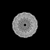
| Biological species |  Autographa californica multiple nucleopolyhedrovirus Autographa californica multiple nucleopolyhedrovirus | |||||||||
| Method | single particle reconstruction / cryo EM / Resolution: 6.3 Å | |||||||||
 Authors Authors | Johnstone BA / Koszalka P / Venugopal H / Coulibaly F | |||||||||
| Funding support |  Australia, 1 items Australia, 1 items
| |||||||||
 Citation Citation | Journal: Sci Adv / Year: 2024 Title: The nucleocapsid architecture and structural atlas of the prototype baculovirus define the hallmarks of a new viral realm. Authors: Bronte A Johnstone / Joshua M Hardy / Jungmin Ha / Anamarija Butkovic / Paulina Koszalka / Cathy Accurso / Hariprasad Venugopal / Alex de Marco / Mart Krupovic / Fasséli Coulibaly /  Abstract: Baculovirus is the most studied insect virus owing to a broad ecological distribution and ease of engineering for biotechnological applications. However, its structure and evolutionary place in the ...Baculovirus is the most studied insect virus owing to a broad ecological distribution and ease of engineering for biotechnological applications. However, its structure and evolutionary place in the virosphere remain enigmatic. Using cryo-electron microscopy, we show that the nucleocapsid forms a covalently cross-linked helical tube protecting a highly compacted 134-kilobase pair DNA genome. The ends of the tube are sealed by the base and cap substructures, which share a 126-subunit hub but differ in components that promote actin tail-mediated propulsion and nuclear entry of the nucleocapsid, respectively. Unexpectedly, sensitive searches for hidden evolutionary links show that the morphogenetic machinery and conserved oral infectivity factors originated within the lineage of baculo-like viruses (class ). The unique viral architecture and structural atlas of hallmark proteins firmly place these viruses into a separate new realm, the highest taxonomy rank, and provide a structural framework to expand their use as sustainable bioinsecticides and biomedical tools. | |||||||||
| History |
|
- Structure visualization
| Supplemental images |
|---|

- Downloads & links
-EMDB archive
| Map data | emd_43590.map.gz | 644 MB |  EMDB map data format EMDB map data format | |
|---|---|---|---|---|
| Header (meta data) | emd-43590-v30.xmlemd-43590.xml | 18.6 KB 18.6 KB | Display Display | EMDB header |
| FSC (resolution estimation) | emd_43590_fsc.xml | 23.4 KB | Display | FSC data file |
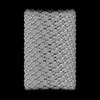
| Images |  emd_43590.png emd_43590.png | 162.5 KB | ||
| Masks | emd_43590_msk_1.map | 1.3 GB | Mask map | |
| Filedesc metadata | emd-43590.cif.gz | 4.8 KB | ||
| Others | emd_43590_additional_1.map.gzemd_43590_half_map_1.map.gzemd_43590_half_map_2.map.gz | 1.2 GB 1.2 GB 1.2 GB | ||
| Archive directory |  http://ftp.pdbj.org/pub/emdb/structures/EMD-43590ftp://ftp.pdbj.org/pub/emdb/structures/EMD-43590 http://ftp.pdbj.org/pub/emdb/structures/EMD-43590ftp://ftp.pdbj.org/pub/emdb/structures/EMD-43590 | HTTPS FTP |
-Related structure data


-Links
| EMDB pages | EMDB (EBI/PDBe) / EMDataResource |
|---|
-Map
| File | Download / File: emd_43590.map.gz / Format: CCP4 / Size: 1.3 GB / Type: IMAGE STORED AS FLOATING POINT NUMBER (4 BYTES) | ||||||||||||||||||||||||||||||||||||
|---|---|---|---|---|---|---|---|---|---|---|---|---|---|---|---|---|---|---|---|---|---|---|---|---|---|---|---|---|---|---|---|---|---|---|---|---|---|
| Projections & slices | Image control
Images are generated by Spider. | ||||||||||||||||||||||||||||||||||||
| Voxel size | X=Y=Z: 1.41 Å | ||||||||||||||||||||||||||||||||||||
| Density |
| ||||||||||||||||||||||||||||||||||||
| Symmetry | Space group: 1 | ||||||||||||||||||||||||||||||||||||
| Details | EMDB XML:
|
 Z (Sec.)
 Y (Row.)
 X (Col.)


-Supplemental data
-Mask #1
| File | emd_43590_msk_1.map | ||||||||||||
|---|---|---|---|---|---|---|---|---|---|---|---|---|---|
| Projections & Slices |
| ||||||||||||
| Density Histograms |


-Additional map: Sharpened map
| File | emd_43590_additional_1.map | ||||||||||||
|---|---|---|---|---|---|---|---|---|---|---|---|---|---|
| Annotation | Sharpened map | ||||||||||||
| Projections & Slices |
| ||||||||||||
| Density Histograms |


-Half map: Half map A
| File | emd_43590_half_map_1.map | ||||||||||||
|---|---|---|---|---|---|---|---|---|---|---|---|---|---|
| Annotation | Half map A | ||||||||||||
| Projections & Slices |
| ||||||||||||
| Density Histograms |


-Half map: Half map B
| File | emd_43590_half_map_2.map | ||||||||||||
|---|---|---|---|---|---|---|---|---|---|---|---|---|---|
| Annotation | Half map B | ||||||||||||
| Projections & Slices |
| ||||||||||||
| Density Histograms |


- Sample components
-Entire : Autographa californica multiple nucleopolyhedrovirus
| Entire | Name: Autographa californica multiple nucleopolyhedrovirus |
|---|---|
| Components |
|
-Supramolecule #1: Autographa californica multiple nucleopolyhedrovirus
| Supramolecule | Name: Autographa californica multiple nucleopolyhedrovirus / type: virus / ID: 1 / Parent: 0 / Macromolecule list: #1 Details: Recombinant AcMNPV (BacToBac, Invitrogen) viruses containing the Bombyx mori cytoplasmic virus polyhedrin gene were amplified in Sf9 cells in suspension culture. NCBI-ID: 307456 Sci species name: Autographa californica multiple nucleopolyhedrovirus Virus type: VIRION / Virus isolate: SPECIES / Virus enveloped: Yes / Virus empty: No |
|---|---|
| Host (natural) | Organism:  Lepidoptera (moths & butterflies) Lepidoptera (moths & butterflies) |
| Virus shell | Shell ID: 1 / Name: Nucleocapsid / Diameter: 500.0 Å |
-Experimental details
-Structure determination
| Method | cryo EM |
|---|---|
 Processing Processing | single particle reconstruction |
| Aggregation state | particle |
-Sample preparation
| Buffer | pH: 7.4 / Details: 10 mM HEPES pH 7.4, 500 mM potassium chloride |
|---|---|
| Grid | Model: Quantifoil R2/2 / Material: GOLD / Mesh: 400 / Pretreatment - Type: GLOW DISCHARGE |
| Vitrification | Cryogen name: ETHANE / Chamber humidity: 100 % / Chamber temperature: 277.15 K / Instrument: FEI VITROBOT MARK IV |
| Details | A suspension of AcMNPV viruses was purified from cell culture supernatant using sucrose gradient ultracentrifugation. Sucrose was removed after purification through dialysis. |
- Electron microscopy
| Microscope | FEI TITAN KRIOS |
|---|---|
| Image recording | Film or detector model: GATAN K3 (6k x 4k) / Number grids imaged: 1 / Number real images: 11439 / Average electron dose: 50.0 e/Å2 |
| Electron beam | Acceleration voltage: 300 kV / Electron source:  FIELD EMISSION GUN FIELD EMISSION GUN |
| Electron optics | Illumination mode: FLOOD BEAM / Imaging mode: BRIGHT FIELD / Cs: 2.7 mm / Nominal defocus max: 1.4000000000000001 µm / Nominal defocus min: 0.8 µm / Nominal magnification: 64000 |
| Experimental equipment |  Model: Titan Krios / Image courtesy: FEI Company |

