Movie
Movie Controller

[English] 日本語
 Yorodumi
- EMDB-43392: Structure of VCP in complex with an ATPase activator and ADP (D2 ... -
+ Open data

- Basic information
| Entry | 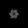 | |||||||||
|---|---|---|---|---|---|---|---|---|---|---|
| Title | Structure of VCP in complex with an ATPase activator and ADP (D2 domains only, hexameric form) | |||||||||
 Map data Map data | ||||||||||
 Sample Sample |
| |||||||||
 Keywords Keywords | activator / complex / ATPase / AAA protein / HYDROLASE / HYDROLASE-ACTIVATOR complex | |||||||||
| Function / homology |  Function and homology information Function and homology informationflavin adenine dinucleotide catabolic process / VCP-NSFL1C complex / endoplasmic reticulum stress-induced pre-emptive quality control / endosome to lysosome transport via multivesicular body sorting pathway / BAT3 complex binding / cytoplasmic ubiquitin ligase complex / cellular response to arsenite ion / protein-DNA covalent cross-linking repair / Derlin-1 retrotranslocation complex / positive regulation of protein K63-linked deubiquitination ...flavin adenine dinucleotide catabolic process / VCP-NSFL1C complex / endoplasmic reticulum stress-induced pre-emptive quality control / endosome to lysosome transport via multivesicular body sorting pathway / BAT3 complex binding / cytoplasmic ubiquitin ligase complex / cellular response to arsenite ion / protein-DNA covalent cross-linking repair / Derlin-1 retrotranslocation complex / positive regulation of protein K63-linked deubiquitination / deubiquitinase activator activity / cytoplasm protein quality control / positive regulation of oxidative phosphorylation / aggresome assembly / ubiquitin-modified protein reader activity / regulation of protein localization to chromatin / mitotic spindle disassembly / VCP-NPL4-UFD1 AAA ATPase complex / cellular response to misfolded protein / positive regulation of mitochondrial membrane potential / vesicle-fusing ATPase / K48-linked polyubiquitin modification-dependent protein binding / NAD+ metabolic process / regulation of aerobic respiration / retrograde protein transport, ER to cytosol / stress granule disassembly / ATPase complex / ubiquitin-specific protease binding / regulation of synapse organization / ciliary transition zone / positive regulation of ATP biosynthetic process / intracellular membrane-bounded organelle / ubiquitin-like protein ligase binding / RHOH GTPase cycle / MHC class I protein binding / autophagosome maturation / negative regulation of hippo signaling / HSF1 activation / endoplasmic reticulum to Golgi vesicle-mediated transport / polyubiquitin modification-dependent protein binding / interstrand cross-link repair / ATP metabolic process / translesion synthesis / Attachment and Entry / negative regulation of protein localization to chromatin / Protein methylation / endoplasmic reticulum unfolded protein response / ERAD pathway / proteasomal protein catabolic process / lipid droplet / ciliary tip / proteasome complex / viral genome replication / Josephin domain DUBs / macroautophagy / negative regulation of smoothened signaling pathway / establishment of protein localization / N-glycan trimming in the ER and Calnexin/Calreticulin cycle / positive regulation of protein-containing complex assembly / Hh mutants are degraded by ERAD / ADP binding / Hedgehog ligand biogenesis / Translesion Synthesis by POLH / Defective CFTR causes cystic fibrosis / positive regulation of non-canonical NF-kappaB signal transduction / ABC-family protein mediated transport / autophagy / cytoplasmic stress granule / Aggrephagy / positive regulation of protein catabolic process / azurophil granule lumen / Ovarian tumor domain proteases / KEAP1-NFE2L2 pathway / positive regulation of canonical Wnt signaling pathway / double-strand break repair / positive regulation of proteasomal ubiquitin-dependent protein catabolic process / cellular response to heat / E3 ubiquitin ligases ubiquitinate target proteins / site of double-strand break / Neddylation / secretory granule lumen / protein phosphatase binding / regulation of apoptotic process / ficolin-1-rich granule lumen / ubiquitin-dependent protein catabolic process / Attachment and Entry / proteasome-mediated ubiquitin-dependent protein catabolic process / ciliary basal body / protein ubiquitination / protein domain specific binding / DNA repair / DNA damage response / Neutrophil degranulation / ubiquitin protein ligase binding / lipid binding / endoplasmic reticulum membrane / perinuclear region of cytoplasm / glutamatergic synapse / endoplasmic reticulum / ATP hydrolysis activity Similarity search - Function | |||||||||
| Biological species |  Homo sapiens (human) Homo sapiens (human) | |||||||||
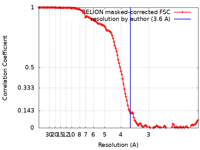
| Method | single particle reconstruction / cryo EM / Resolution: 3.6 Å | |||||||||
 Authors Authors | Jones NH / Urnivicius L / Kapoor TM | |||||||||
| Funding support |  United States, 1 items United States, 1 items
| |||||||||
 Citation Citation | Journal: bioRxiv / Year: 2023 Title: Allosteric activation of VCP, a AAA unfoldase, by small molecule mimicry. Authors: N H Jones / Q Liu / L Urnavicius / N E Dahan / L E Vostal / T M Kapoor Abstract: The loss of function of AAA (ATPases associated with diverse cellular activities) mechanoenzymes has been linked to diseases, and small molecules that activate these proteins can be powerful tools to ...The loss of function of AAA (ATPases associated with diverse cellular activities) mechanoenzymes has been linked to diseases, and small molecules that activate these proteins can be powerful tools to probe mechanisms and test therapeutic hypotheses. Unlike chemical inhibitors that can bind a single conformational state to block enzyme activity, activator binding must be permissive to different conformational states needed for enzyme function. However, we do not know how AAA proteins can be activated by small molecules. Here, we focus on valosin-containing protein (VCP)/p97, a AAA unfoldase whose loss of function has been linked to protein aggregation-based disorders, to identify druggable sites for chemical activators. We identified VCP Activator 1 (VA1), a compound that dose-dependently stimulates VCP ATPase activity up to ∼3-fold. Our cryo-EM studies resulted in structures (∼2.9-3.5 Å-resolution) of VCP in apo and ADP-bound states, and revealed VA1 binding an allosteric pocket near the C-terminus in both states. Engineered mutations in the VA1 binding site confer resistance to VA1, and furthermore, modulate VCP activity to a similar level as VA1-mediated activation. The VA1 binding site can alternatively be occupied by a phenylalanine residue in the VCP C-terminal tail, a motif that is post-translationally modified and interacts with cofactors. Together, our findings uncover a druggable allosteric site and a mechanism of enzyme regulation that can be tuned through small molecule mimicry. SIGNIFICANCE: The loss of function of valosin-containing protein (VCP/p97), a mechanoenzyme from the AAA superfamily that hydrolyzes ATP and uses the released energy to extract or unfold substrate ...SIGNIFICANCE: The loss of function of valosin-containing protein (VCP/p97), a mechanoenzyme from the AAA superfamily that hydrolyzes ATP and uses the released energy to extract or unfold substrate proteins, is linked to protein aggregation-based disorders. However, druggable allosteric sites to activate VCP, or any AAA mechanoenzyme, have not been identified. Here, we report cryo-EM structures of VCP in two states in complex with VA1, a compound we identified that dose-dependently stimulates VCP's ATP hydrolysis activity. The VA1 binding site can also be occupied by a phenylalanine residue in the VCP C-terminal tail, suggesting that VA1 acts through mimicry of this interaction. Our study reveals a druggable allosteric site and a mechanism of enzyme regulation. | |||||||||
| History |
|
- Structure visualization
| Supplemental images |
|---|

- Downloads & links
-EMDB archive
| Map data | emd_43392.map.gz | 177.9 MB | EMDB map data format | |
|---|---|---|---|---|
| Header (meta data) | emd-43392-v30.xmlemd-43392.xml | 19.6 KB 19.6 KB | Display Display | EMDB header |
| FSC (resolution estimation) | emd_43392_fsc.xml | 13 KB | Display | FSC data file |
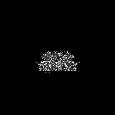
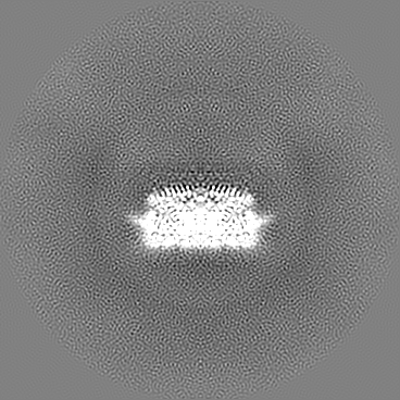
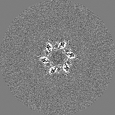
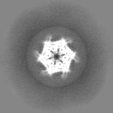
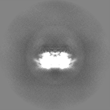
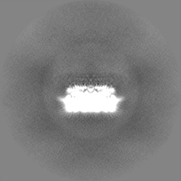
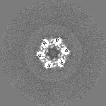
| Images | 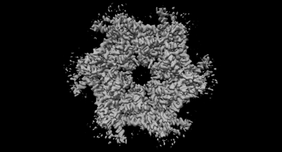 emd_43392.png emd_43392.png | 54.6 KB | ||
| Filedesc metadata | emd-43392.cif.gz | 6.7 KB | ||
| Others | emd_43392_half_map_1.map.gzemd_43392_half_map_2.map.gz | 150 MB 150.3 MB | ||
| Archive directory |  http://ftp.pdbj.org/pub/emdb/structures/EMD-43392ftp://ftp.pdbj.org/pub/emdb/structures/EMD-43392 http://ftp.pdbj.org/pub/emdb/structures/EMD-43392ftp://ftp.pdbj.org/pub/emdb/structures/EMD-43392 | HTTPS FTP |
-Related structure data
| Related structure data | 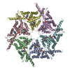 8vovMC 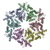 8vkuC 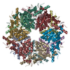 8vlsC C: citing same article ( M: atomic model generated by this map |
|---|---|
| Similar structure data |


-Links
| EMDB pages | EMDB (EBI/PDBe) / EMDataResource |
|---|---|
| Related items in Molecule of the Month |


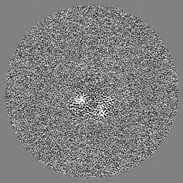
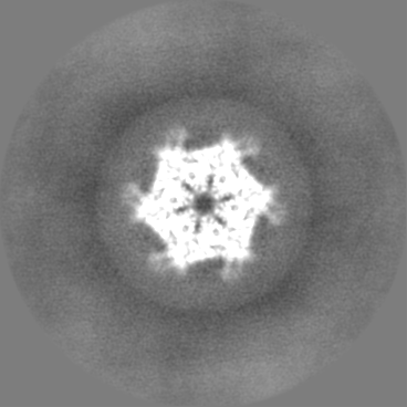
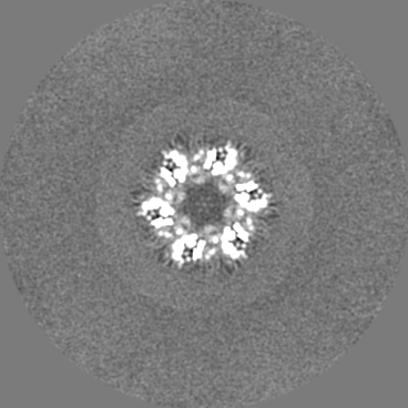
-Map
| File | Download / File: emd_43392.map.gz / Format: CCP4 / Size: 190.1 MB / Type: IMAGE STORED AS FLOATING POINT NUMBER (4 BYTES) | ||||||||||||||||||||||||||||||||||||
|---|---|---|---|---|---|---|---|---|---|---|---|---|---|---|---|---|---|---|---|---|---|---|---|---|---|---|---|---|---|---|---|---|---|---|---|---|---|
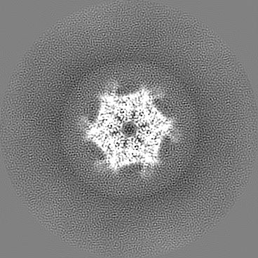
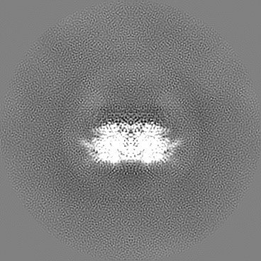
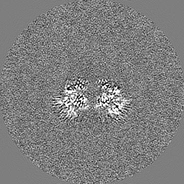
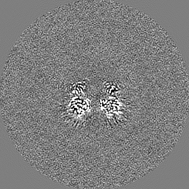
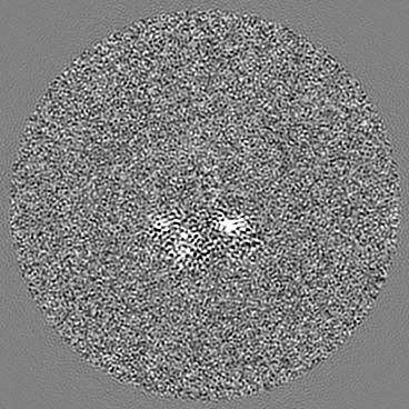
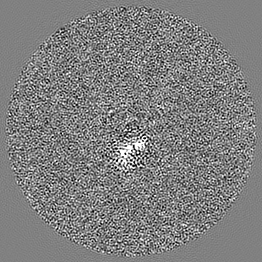
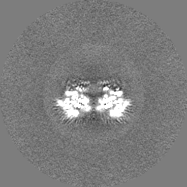
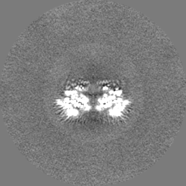
| Projections & slices | Image control
Images are generated by Spider. | ||||||||||||||||||||||||||||||||||||
| Voxel size | X=Y=Z: 1.035 Å | ||||||||||||||||||||||||||||||||||||
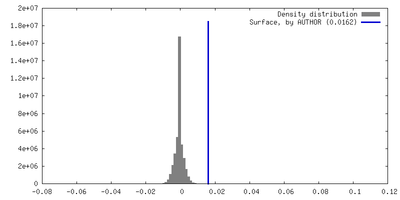
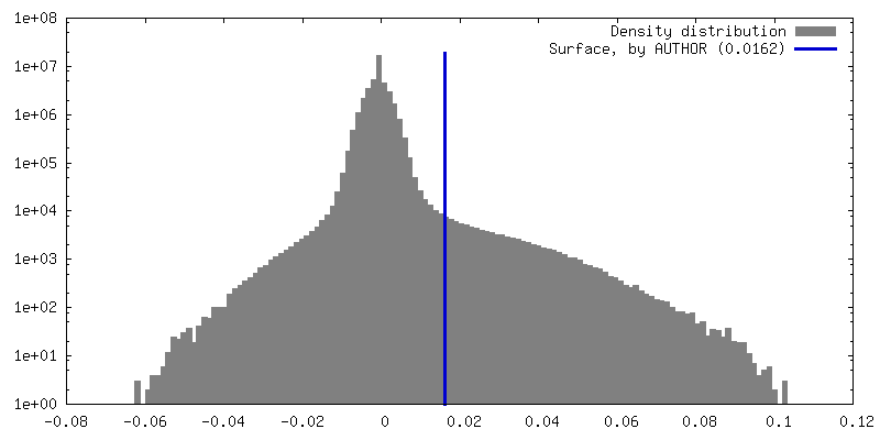
| Density |
| ||||||||||||||||||||||||||||||||||||
| Symmetry | Space group: 1 | ||||||||||||||||||||||||||||||||||||
| Details | EMDB XML:
|
 Z (Sec.)
 Y (Row.)
 X (Col.)


-Supplemental data
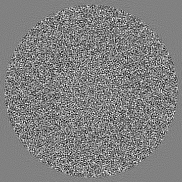
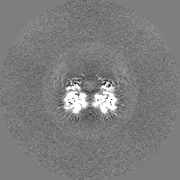
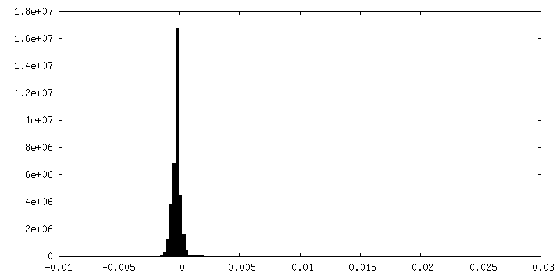
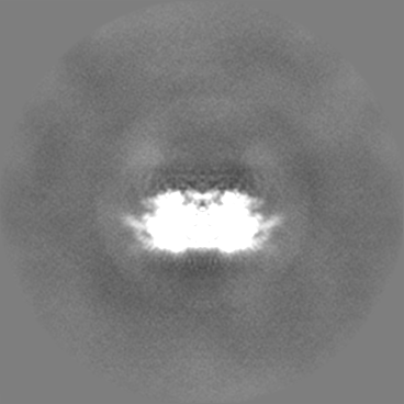
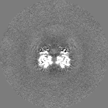
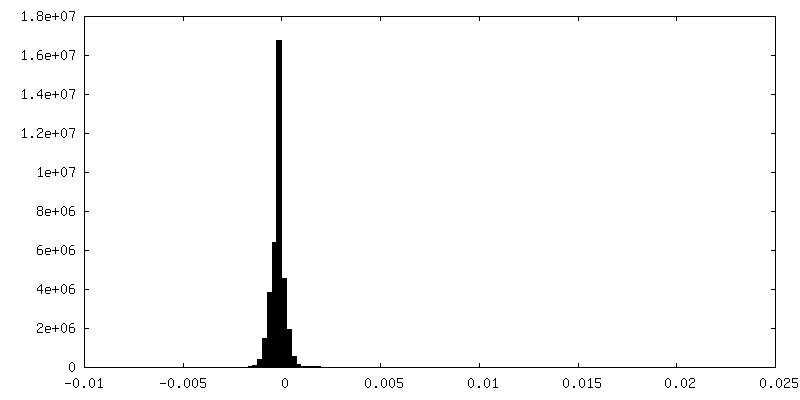
-Half map: #1
| File | emd_43392_half_map_1.map | ||||||||||||
|---|---|---|---|---|---|---|---|---|---|---|---|---|---|
| Projections & Slices |
| ||||||||||||
| Density Histograms |
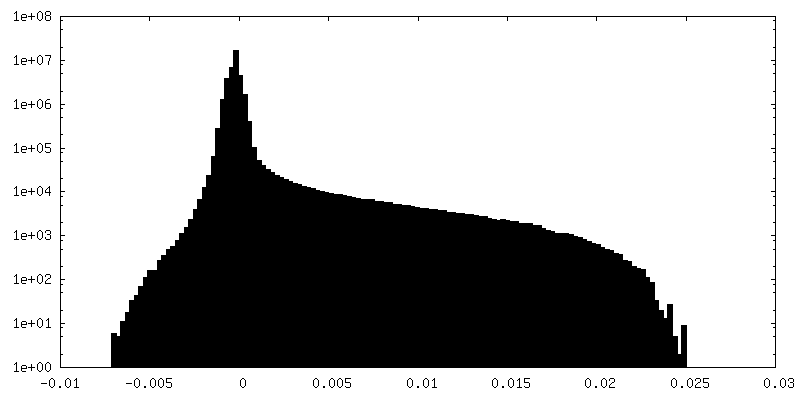
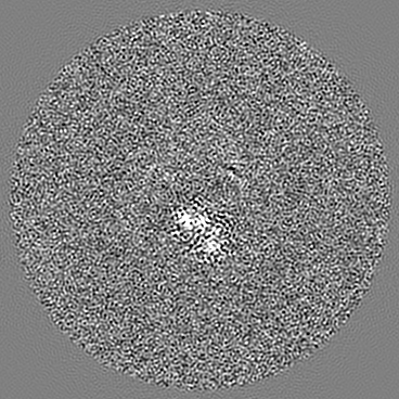
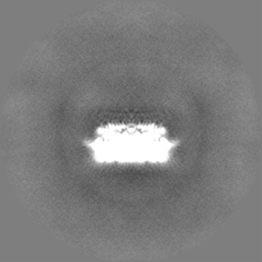
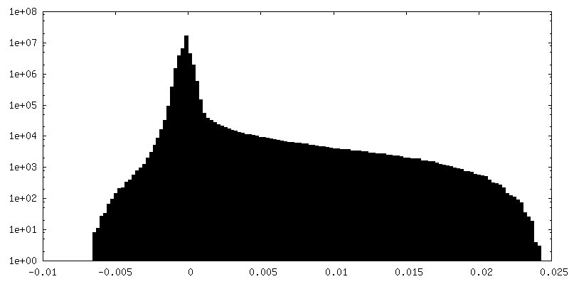
-Half map: #2
| File | emd_43392_half_map_2.map | ||||||||||||
|---|---|---|---|---|---|---|---|---|---|---|---|---|---|
| Projections & Slices |
| ||||||||||||
| Density Histograms |
- Sample components
-Entire : Complex of VCP with ADP and ATPase activator small molecule VAA1
| Entire | Name: Complex of VCP with ADP and ATPase activator small molecule VAA1 |
|---|---|
| Components |
|
-Supramolecule #1: Complex of VCP with ADP and ATPase activator small molecule VAA1
| Supramolecule | Name: Complex of VCP with ADP and ATPase activator small molecule VAA1 type: complex / ID: 1 / Parent: 0 / Macromolecule list: #1 |
|---|---|
| Source (natural) | Organism: Homo sapiens (human) |
-Macromolecule #1: Transitional endoplasmic reticulum ATPase
| Macromolecule | Name: Transitional endoplasmic reticulum ATPase / type: protein_or_peptide / ID: 1 / Number of copies: 6 / Enantiomer: LEVO / EC number: vesicle-fusing ATPase |
|---|---|
| Source (natural) | Organism: Homo sapiens (human) |
| Molecular weight | Theoretical: 89.43682 KDa |
| Recombinant expression | Organism:  |
| Sequence | String: MASGADSKGD DLSTAILKQK NRPNRLIVDE AINEDNSVVS LSQPKMDELQ LFRGDTVLLK GKKRREAVCI VLSDDTCSDE KIRMNRVVR NNLRVRLGDV ISIQPCPDVK YGKRIHVLPI DDTVEGITGN LFEVYLKPYF LEAYRPIRKG DIFLVRGGMR A VEFKVVET ...String: MASGADSKGD DLSTAILKQK NRPNRLIVDE AINEDNSVVS LSQPKMDELQ LFRGDTVLLK GKKRREAVCI VLSDDTCSDE KIRMNRVVR NNLRVRLGDV ISIQPCPDVK YGKRIHVLPI DDTVEGITGN LFEVYLKPYF LEAYRPIRKG DIFLVRGGMR A VEFKVVET DPSPYCIVAP DTVIHCEGEP IKREDEEESL NEVGYDDIGG CRKQLAQIKE MVELPLRHPA LFKAIGVKPP RG ILLYGPP GTGKTLIARA VANETGAFFF LINGPEIMSK LAGESESNLR KAFEEAEKNA PAIIFIDELD AIAPKREKTH GEV ERRIVS QLLTLMDGLK QRAHVIVMAA TNRPNSIDPA LRRFGRFDRE VDIGIPDATG RLEILQIHTK NMKLADDVDL EQVA NETHG HVGADLAALC SEAALQAIRK KMDLIDLEDE TIDAEVMNSL AVTMDDFRWA LSQSNPSALR ETVVEVPQVT WEDIG GLED VKRELQELVQ YPVEHPDKFL KFGMTPSKGV LFYGPPGCGK TLLAKAIANE CQANFISIKG PELLTMWFGE SEANVR EIF DKARQAAPCV LFFDELDSIA KARGGNIGDG GGAADRVINQ ILTEMDGMST KKNVFIIGAT NRPDIIDPAI LRPGRLD QL IYIPLPDEKS RVAILKANLR KSPVAKDVDL EFLAKMTNGF SGADLTEICQ RACKLAIRES IESEIRRERE RQTNPSAM E VEEDDPVPEI RRDHFEEAMR FARRSVSDND IRKYEMFAQT LQQSRGFGSF RFPSGNQGGA GPSQGSGGGT GGSVYTEDN DDDLYG UniProtKB: Transitional endoplasmic reticulum ATPase |
-Macromolecule #2: ADENOSINE-5'-DIPHOSPHATE
| Macromolecule | Name: ADENOSINE-5'-DIPHOSPHATE / type: ligand / ID: 2 / Number of copies: 6 / Formula: ADP |
|---|---|
| Molecular weight | Theoretical: 427.201 Da |
| Chemical component information |  ChemComp-ADP: |
-Macromolecule #3: (3R)-N-[2-(ethylsulfanyl)phenyl]-3-(1-oxo-1,3-dihydro-2H-isoindol...
| Macromolecule | Name: (3R)-N-[2-(ethylsulfanyl)phenyl]-3-(1-oxo-1,3-dihydro-2H-isoindol-2-yl)butanamide type: ligand / ID: 3 / Number of copies: 6 / Formula: A1AC1 |
|---|---|
| Molecular weight | Theoretical: 354.466 Da |
-Experimental details
-Structure determination
| Method | cryo EM |
|---|---|
 Processing Processing | single particle reconstruction |
| Aggregation state | particle |
-Sample preparation
| Concentration | 1 mg/mL | |||||||||||||||||||||
|---|---|---|---|---|---|---|---|---|---|---|---|---|---|---|---|---|---|---|---|---|---|---|
| Buffer | pH: 7.5 Component:
Details: 50 mM K.HEPES pH 7.5, 25 mM KCl, 2.5 mM MgCl2, 2.5 mM GSH, 0.5% DMSO, 0.01% FOM | |||||||||||||||||||||
| Grid | Model: Quantifoil R2/2 / Material: COPPER / Mesh: 300 / Pretreatment - Type: GLOW DISCHARGE / Pretreatment - Time: 30 sec. | |||||||||||||||||||||
| Vitrification | Cryogen name: ETHANE / Chamber humidity: 100 % / Chamber temperature: 298 K / Instrument: FEI VITROBOT MARK IV |
- Electron microscopy
| Microscope | FEI TITAN KRIOS |
|---|---|
| Image recording | Film or detector model: GATAN K3 (6k x 4k) / Average electron dose: 38.3 e/Å2 |
| Electron beam | Acceleration voltage: 300 kV / Electron source:  FIELD EMISSION GUN FIELD EMISSION GUN |
| Electron optics | Illumination mode: FLOOD BEAM / Imaging mode: BRIGHT FIELD / Nominal defocus max: 3.5 µm / Nominal defocus min: 0.5 µm |
| Experimental equipment |  Model: Titan Krios / Image courtesy: FEI Company |