National Institutes of Health/National Heart, Lung, and Blood Institute (NIH/NHLBI)
R01HL145473
米国
引用
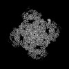
ジャーナル: Proc Natl Acad Sci U S A / 年: 2024 タイトル: Structural insights into the regulation of RyR1 by S100A1. 著者: Gunnar Weninger / Marco C Miotto / Carl Tchagou / Steven Reiken / Haikel Dridi / Sören Brandenburg / Gabriel C Riedemann / Qi Yuan / Yang Liu / Alexander Chang / Anetta Wronska / Stephan E ...著者: Gunnar Weninger / Marco C Miotto / Carl Tchagou / Steven Reiken / Haikel Dridi / Sören Brandenburg / Gabriel C Riedemann / Qi Yuan / Yang Liu / Alexander Chang / Anetta Wronska / Stephan E Lehnart / Andrew R Marks / 要旨: S100A1, a small homodimeric EF-hand Ca-binding protein (~21 kDa), plays an important regulatory role in Ca signaling pathways involved in various biological functions including Ca cycling and ...S100A1, a small homodimeric EF-hand Ca-binding protein (~21 kDa), plays an important regulatory role in Ca signaling pathways involved in various biological functions including Ca cycling and contractile performance in skeletal and cardiac myocytes. One key target of the S100A1 interactome is the ryanodine receptor (RyR), a huge homotetrameric Ca release channel (~2.3 MDa) of the sarcoplasmic reticulum. Here, we report cryoelectron microscopy structures of S100A1 bound to RyR1, the skeletal muscle isoform, in absence and presence of Ca. Ca-free apo-S100A1 binds beneath the bridging solenoid (BSol) and forms contacts with the junctional solenoid and the shell-core linker of RyR1. Upon Ca-binding, S100A1 undergoes a conformational change resulting in the exposure of the hydrophobic pocket known to serve as a major interaction site of S100A1. Through interactions of the hydrophobic pocket with RyR1, Ca-bound S100A1 intrudes deeper into the RyR1 structure beneath BSol than the apo-form and induces sideways motions of the C-terminal BSol region toward the adjacent RyR1 protomer resulting in tighter interprotomer contacts. Interestingly, the second hydrophobic pocket of the S100A1-dimer is largely exposed at the hydrophilic surface making it prone to interactions with the local environment, suggesting that S100A1 could be involved in forming larger heterocomplexes of RyRs with other protein partners. Since S100A1 interactions stabilizing BSol are implicated in the regulation of RyR-mediated Ca release, the characterization of the S100A1 binding site conserved between RyR isoforms may provide the structural basis for the development of therapeutic strategies regarding treatments of RyR-related disorders.
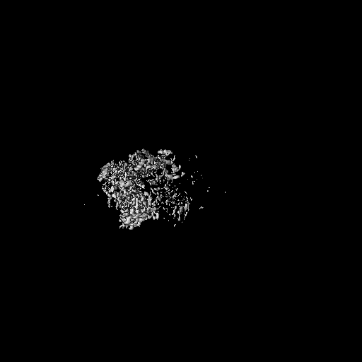
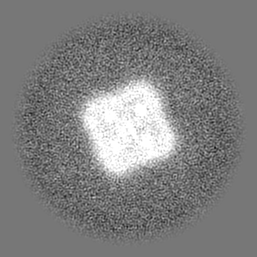
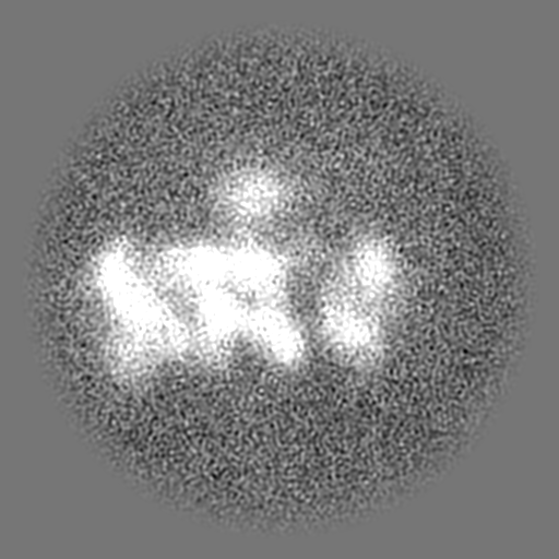
全体 : Complex of RyR1 with Calstabin-1 and S100A1 (high-Ca2+/CFF/ATP co...
全体
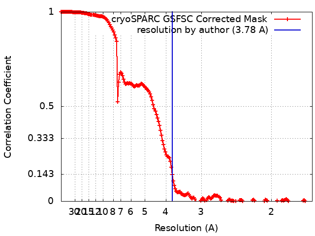
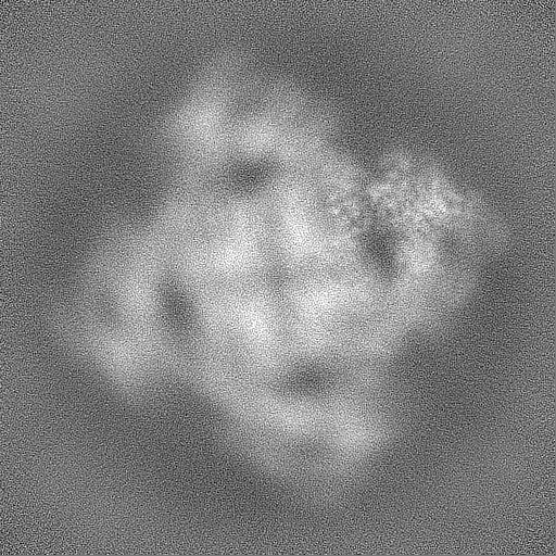
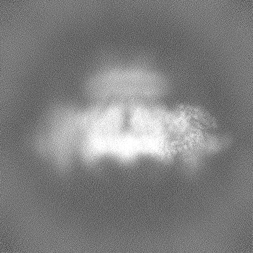
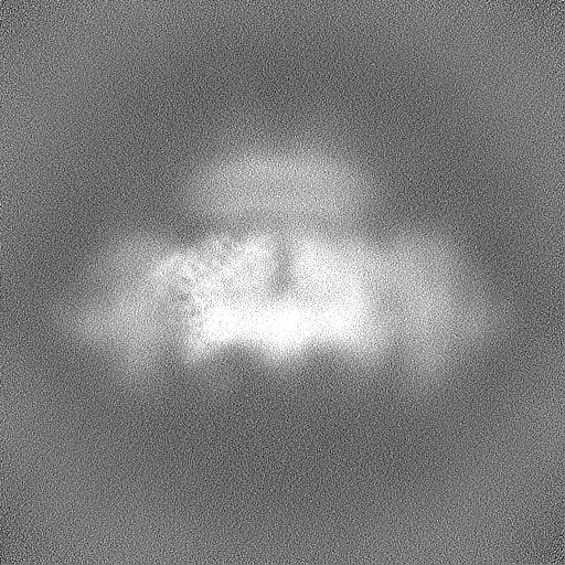
名称: Complex of RyR1 with Calstabin-1 and S100A1 (high-Ca2+/CFF/ATP condition)
要素
複合体: Complex of RyR1 with Calstabin-1 and S100A1 (high-Ca2+/CFF/ATP condition)
-
超分子 #1: Complex of RyR1 with Calstabin-1 and S100A1 (high-Ca2+/CFF/ATP co...
超分子
名称: Complex of RyR1 with Calstabin-1 and S100A1 (high-Ca2+/CFF/ATP condition) タイプ: complex / ID: 1 / 親要素: 0 / 含まれる分子: #1-#3 / 詳細: 0.25 mM free Ca2+; 5 mM Caffeine; 10 mM ATP
由来(天然)
生物種: Mus musculus (ハツカネズミ)
-
実験情報
-
構造解析
手法
クライオ電子顕微鏡法
解析
単粒子再構成法
試料の集合状態
particle
-
試料調製
濃度
8.5 mg/mL
緩衝液
pH: 7.4 構成要素:
濃度
名称
式
10.0 mmol/L
HEPES
150.0 mmol/L
sodium chloride
NaCl
1.0 mmol/L
EGTA
0.25 %
CHAPS
0.01 %
DOPC
0.5 mmol/L
TCEP
グリッド
モデル: Quantifoil R0.6/1 / 材質: GOLD / メッシュ: 300
凍結
凍結剤: ETHANE / チャンバー内湿度: 100 % / チャンバー内温度: 277.15 K / 装置: FEI VITROBOT MARK IV
 ムービー
ムービー コントローラー


 データを開く
 基本情報
基本情報
 マップデータ
 試料
試料 キーワード
キーワード
 データ登録者
 米国, 1件
  引用
引用
 構造の表示
構造の表示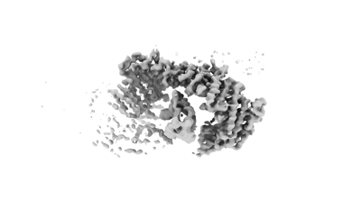
 ダウンロードとリンク
 EMDBマップデータ形式
 emd_43302.png
 http://ftp.pdbj.org/pub/emdb/structures/EMD-43302


 Z (Sec.)
 Y (Row.)
 X (Col.)


 試料の構成要素
 解析
解析 電子顕微鏡法
 FIELD EMISSION GUN