 Movie
Movie Controller

[English] 日本語
 Yorodumi
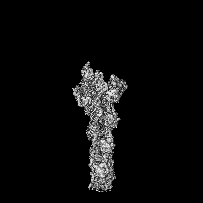
- EMDB-43274: Structure of full-length gelsolin bound to the barbed end of F-actin -
+ Open data

- Basic information
| Entry |  | |||||||||
|---|---|---|---|---|---|---|---|---|---|---|
| Title | Structure of full-length gelsolin bound to the barbed end of F-actin | |||||||||
 Map data Map data | ||||||||||
 Sample Sample |
| |||||||||
 Keywords Keywords | cytoskeleton / actin / cell motility / PROTEIN BINDING | |||||||||
| Function / homology |  Function and homology information Function and homology informationstriated muscle atrophy / regulation of establishment of T cell polarity / regulation of plasma membrane raft polarization / regulation of receptor clustering / positive regulation of keratinocyte apoptotic process / renal protein absorption / positive regulation of protein processing in phagocytic vesicle / phosphatidylinositol 3-kinase catalytic subunit binding / positive regulation of actin nucleation / actin cap ...striated muscle atrophy / regulation of establishment of T cell polarity / regulation of plasma membrane raft polarization / regulation of receptor clustering / positive regulation of keratinocyte apoptotic process / renal protein absorption / positive regulation of protein processing in phagocytic vesicle / phosphatidylinositol 3-kinase catalytic subunit binding / positive regulation of actin nucleation / actin cap / myosin II binding / host-mediated suppression of symbiont invasion / cell projection assembly / actin filament severing / barbed-end actin filament capping / actin polymerization or depolymerization / actin filament depolymerization / actin filament capping / relaxation of cardiac muscle / Sensory processing of sound by outer hair cells of the cochlea / phagocytosis, engulfment / hepatocyte apoptotic process / cardiac muscle cell contraction / cytoskeletal motor activator activity / myosin heavy chain binding / tropomyosin binding / actin filament bundle / troponin I binding / filamentous actin / mesenchyme migration / skeletal muscle myofibril / sarcoplasm / striated muscle thin filament / actin filament bundle assembly / skeletal muscle thin filament assembly / actin monomer binding / Caspase-mediated cleavage of cytoskeletal proteins / cilium assembly / response to muscle stretch / skeletal muscle fiber development / actin filament polymerization / stress fiber / phagocytic vesicle / titin binding / phosphatidylinositol-4,5-bisphosphate binding / actin filament organization / central nervous system development / actin filament / filopodium / protein destabilization / Hydrolases; Acting on acid anhydrides; Acting on acid anhydrides to facilitate cellular and subcellular movement / calcium-dependent protein binding / actin filament binding / actin cytoskeleton / lamellipodium / actin binding / cell body / secretory granule lumen / blood microparticle / amyloid fibril formation / ficolin-1-rich granule lumen / Amyloid fiber formation / protein domain specific binding / focal adhesion / hydrolase activity / positive regulation of gene expression / calcium ion binding / Neutrophil degranulation / magnesium ion binding / : / extracellular exosome / extracellular region / ATP binding / identical protein binding / plasma membrane / cytosol / cytoplasm Similarity search - Function | |||||||||
| Biological species |   Homo sapiens (human) Homo sapiens (human) | |||||||||
| Method | single particle reconstruction / cryo EM / Resolution: 2.63 Å | |||||||||
 Authors Authors | Barrie KR / Rebowski G / Dominguez R | |||||||||
| Funding support |  United States, 1 items United States, 1 items
| |||||||||
 Citation Citation | Journal: bioRxiv / Year: 2024 Title: Mechanism of Actin Filament Severing and Capping by Gelsolin. Authors: Kyle R Barrie / Grzegorz Rebowski / Roberto Dominguez Abstract: Gelsolin is the prototypical member of a family of Ca -dependent F-actin severing and capping proteins. A structure of Ca -bound full-length gelsolin at the barbed end shows domains G1G6 and the ...Gelsolin is the prototypical member of a family of Ca -dependent F-actin severing and capping proteins. A structure of Ca -bound full-length gelsolin at the barbed end shows domains G1G6 and the inter-domain linkers wrapping around F-actin. Another structure shows domains G1G3, a fragment produced during apoptosis, on both sides of F-actin. Conformational changes that trigger severing occur on one side of F-actin with G1G6 and on both sides with G1G3. Gelsolin remains bound after severing, blocking subunit exchange. | |||||||||
| History |
|
- Structure visualization
| Supplemental images |
|---|

- Downloads & links
-EMDB archive
| Map data | emd_43274.map.gz | 131.2 MB | EMDB map data format | |
|---|---|---|---|---|
| Header (meta data) | emd-43274-v30.xmlemd-43274.xml | 17.1 KB 17.1 KB | Display Display | EMDB header |
| Images |  emd_43274.png emd_43274.png | 52.9 KB | ||
| Filedesc metadata | emd-43274.cif.gz | 7 KB | ||
| Archive directory |  http://ftp.pdbj.org/pub/emdb/structures/EMD-43274ftp://ftp.pdbj.org/pub/emdb/structures/EMD-43274 http://ftp.pdbj.org/pub/emdb/structures/EMD-43274ftp://ftp.pdbj.org/pub/emdb/structures/EMD-43274 | HTTPS FTP |
-Related structure data
| Related structure data |  8vizMC  8vkhC C: citing same article ( M: atomic model generated by this map |
|---|---|
| Similar structure data |


-Links
| EMDB pages | EMDB (EBI/PDBe) / EMDataResource |
|---|---|
| Related items in Molecule of the Month |


-Map
| File | Download / File: emd_43274.map.gz / Format: CCP4 / Size: 274.6 MB / Type: IMAGE STORED AS FLOATING POINT NUMBER (4 BYTES) | ||||||||||||||||||||||||||||||||||||
|---|---|---|---|---|---|---|---|---|---|---|---|---|---|---|---|---|---|---|---|---|---|---|---|---|---|---|---|---|---|---|---|---|---|---|---|---|---|
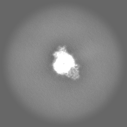
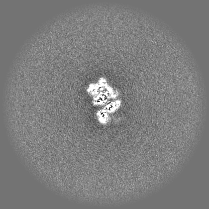
| Projections & slices | Image control
Images are generated by Spider. | ||||||||||||||||||||||||||||||||||||
| Voxel size | X=Y=Z: 1.08 Å | ||||||||||||||||||||||||||||||||||||
| Density |
| ||||||||||||||||||||||||||||||||||||
| Symmetry | Space group: 1 | ||||||||||||||||||||||||||||||||||||
| Details | EMDB XML:
|
 Z (Sec.)
 Y (Row.)
 X (Col.)


-Supplemental data
- Sample components
-Entire : Structure of full-length cytoplasmic gelsolin bound to the barbed...
| Entire | Name: Structure of full-length cytoplasmic gelsolin bound to the barbed end of F-actin |
|---|---|
| Components |
|
-Supramolecule #1: Structure of full-length cytoplasmic gelsolin bound to the barbed...
| Supramolecule | Name: Structure of full-length cytoplasmic gelsolin bound to the barbed end of F-actin type: complex / ID: 1 / Parent: 0 / Macromolecule list: #1-#2 |
|---|---|
| Source (natural) | Organism: |
-Supramolecule #2: skeletal actin
| Supramolecule | Name: skeletal actin / type: complex / ID: 2 / Parent: 1 |
|---|---|
| Source (natural) | Organism: |
-Macromolecule #1: Actin, alpha skeletal muscle
| Macromolecule | Name: Actin, alpha skeletal muscle / type: protein_or_peptide / ID: 1 / Number of copies: 6 / Enantiomer: LEVO |
|---|---|
| Source (natural) | Organism: |
| Molecular weight | Theoretical: 41.875633 KDa |
| Sequence | String: DEDETTALVC DNGSGLVKAG FAGDDAPRAV FPSIVGRPRH QGVMVGMGQK DSYVGDEAQS KRGILTLKYP IE(HIC)GII TNW DDMEKIWHHT FYNELRVAPE EHPTLLTEAP LNPKANREKM TQIMFETFNV PAMYVAIQAV LSLYASGRTT GIVLDSG DG VTHNVPIYEG ...String: DEDETTALVC DNGSGLVKAG FAGDDAPRAV FPSIVGRPRH QGVMVGMGQK DSYVGDEAQS KRGILTLKYP IE(HIC)GII TNW DDMEKIWHHT FYNELRVAPE EHPTLLTEAP LNPKANREKM TQIMFETFNV PAMYVAIQAV LSLYASGRTT GIVLDSG DG VTHNVPIYEG YALPHAIMRL DLAGRDLTDY LMKILTERGY SFVTTAEREI VRDIKEKLCY VALDFENEMA TAASSSSL E KSYELPDGQV ITIGNERFRC PETLFQPSFI GMESAGIHET TYNSIMKCDI DIRKDLYANN VMSGGTTMYP GIADRMQKE ITALAPSTMK IKIIAPPERK YSVWIGGSIL ASLSTFQQMW ITKQEYDEAG PSIVHRKCF UniProtKB: Actin, alpha skeletal muscle |
-Macromolecule #2: Gelsolin
| Macromolecule | Name: Gelsolin / type: protein_or_peptide / ID: 2 / Number of copies: 1 / Enantiomer: LEVO |
|---|---|
| Source (natural) | Organism: Homo sapiens (human) |
| Molecular weight | Theoretical: 82.73768 KDa |
| Recombinant expression | Organism:  |
| Sequence | String: MHHHHHHMVV EHPEFLKAGK EPGLQIWRVE KFDLVPVPTN LYGDFFTGDA YVILKTVQLR NGNLQYDLHY WLGNECSQDE SGAAAIFTV QLDDYLNGRA VQHREVQGFE SATFLGYFKS GLKYKKGGVA SGFKHVVPNE VVVQRLFQVK GRRVVRATEV P VSWESFNN ...String: MHHHHHHMVV EHPEFLKAGK EPGLQIWRVE KFDLVPVPTN LYGDFFTGDA YVILKTVQLR NGNLQYDLHY WLGNECSQDE SGAAAIFTV QLDDYLNGRA VQHREVQGFE SATFLGYFKS GLKYKKGGVA SGFKHVVPNE VVVQRLFQVK GRRVVRATEV P VSWESFNN GDCFILDLGN NIHQWCGSNS NRYERLKATQ VSKGIRDNER SGRARVHVSE EGTEPEAMLQ VLGPKPALPA GT EDTAKED AANRKLAKLY KVSNGAGTMS VSLVADENPF AQGALKSEDC FILDHGKDGK IFVWKGKQAN TEERKAALKT ASD FITKMD YPKQTQVSVL PEGGETPLFK QFFKNWRDPD QTDGLGLSYL SSHIANVERV PFDAATLHTS TAMAAQHGMD DDGT GQKQI WRIEGSNKVP VDPATYGQFY GGDSYIILYN YRHGGRQGQI IYNWQGAQST QDEVAASAIL TAQLDEELGG TPVQS RVVQ GKEPAHLMSL FGGKPMIIYK GGTSREGGQT APASTRLFQV RANSAGATRA VEVLPKAGAL NSNDAFVLKT PSAAYL WVG TGASEAEKTG AQELLRVLRA QPVQVAEGSE PDGFWEALGG KAAYRTSPRL KDKKMDAHPP RLFACSNKIG RFVIEEV PG ELMQEDLATD DVMLLDTWDQ VFVWVGKDSQ EEEKTEALTS AKRYIETDPA NRDRRTPITV VKQGFEPPSF VGWFLGWD D DYWSVDPLDR AMAELAAWSH PQFEK UniProtKB: Gelsolin |
-Macromolecule #3: ADENOSINE-5'-DIPHOSPHATE
| Macromolecule | Name: ADENOSINE-5'-DIPHOSPHATE / type: ligand / ID: 3 / Number of copies: 5 / Formula: ADP |
|---|---|
| Molecular weight | Theoretical: 427.201 Da |
| Chemical component information |  ChemComp-ADP: |
-Macromolecule #4: CALCIUM ION
| Macromolecule | Name: CALCIUM ION / type: ligand / ID: 4 / Number of copies: 14 / Formula: CA |
|---|---|
| Molecular weight | Theoretical: 40.078 Da |
-Macromolecule #5: ADENOSINE-5'-TRIPHOSPHATE
| Macromolecule | Name: ADENOSINE-5'-TRIPHOSPHATE / type: ligand / ID: 5 / Number of copies: 1 / Formula: ATP |
|---|---|
| Molecular weight | Theoretical: 507.181 Da |
| Chemical component information |  ChemComp-ATP: |
-Experimental details
-Structure determination
| Method | cryo EM |
|---|---|
 Processing Processing | single particle reconstruction |
| Aggregation state | particle |
-Sample preparation
| Buffer | pH: 7.5 |
|---|---|
| Vitrification | Cryogen name: ETHANE / Instrument: FEI VITROBOT MARK IV |
- Electron microscopy
| Microscope | FEI TITAN KRIOS |
|---|---|
| Image recording | Film or detector model: GATAN K3 (6k x 4k) / Average electron dose: 45.0 e/Å2 |
| Electron beam | Acceleration voltage: 300 kV / Electron source: OTHER |
| Electron optics | Illumination mode: OTHER / Imaging mode: OTHER / Nominal defocus max: 2.5 µm / Nominal defocus min: 0.5 µm |
| Experimental equipment |  Model: Titan Krios / Image courtesy: FEI Company |
