National Institutes of Health/National Institute of General Medical Sciences (NIH/NIGMS)
R01 GM125844
United States
National Institutes of Health/National Institute of General Medical Sciences (NIH/NIGMS)
R01 GM108753
United States
National Institutes of Health/National Institute of General Medical Sciences (NIH/NIGMS)
R01 GM143805
United States
National Institutes of Health/National Institute of General Medical Sciences (NIH/NIGMS)
F31 GM139324
United States
Citation
Journal: J Mol Biol / Year: 2025 Title: The Structure of the Drp1 Lattice on Membrane. Authors: Ruizhi Peng / Kristy Rochon / Anelise N Hutson / Scott M Stagg / Jason A Mears / Abstract: Mitochondrial health relies on the membrane fission mediated by dynamin-related protein 1 (Drp1). Previous structural studies of Drp1 on remodeled membranes were hampered by heterogeneity, leaving a ...Mitochondrial health relies on the membrane fission mediated by dynamin-related protein 1 (Drp1). Previous structural studies of Drp1 on remodeled membranes were hampered by heterogeneity, leaving a critical gap in the understanding of the mitochondrial fission mechanisms. Here we present a cryo-electron microscopy structure of full-length human Drp1 decorated on membrane tubules. Using the reconstruction of average subtracted tubular regions (RASTR) technique, we report that Drp1 forms a locally ordered lattice along the tubule without global helical symmetry. The filaments in the lattice are similar to dynamin rungs with conserved stalk interactions. Adjacent filaments are connected by GTPase domain interactions in a novel stacked conformation. We identified two states of the Drp1 lattice among the heterogenous dataset representing conformational changes around hinge 1. Additionally, we observed contact between Drp1 and membrane that can be assigned to the variable domain sequence. Together these structures revealed a putative mechanism by which Drp1 constricts mitochondria membranes in a stepwise, "ratchet" manner.
Details: Primary Buffer: 25 mM HEPES (KOH) pH 7.5, 0.15 M KCl, 5 mM MgCl2, 10 mM Beta-mercaptoethanol Nanotubes: 40% D-galactosyl-beta-1-N-nervonyl-erythro-sphingosine (GC), 35% ...Details: Primary Buffer: 25 mM HEPES (KOH) pH 7.5, 0.15 M KCl, 5 mM MgCl2, 10 mM Beta-mercaptoethanol Nanotubes: 40% D-galactosyl-beta-1-N-nervonyl-erythro-sphingosine (GC), 35% phosphatidylethanolamine (PE), 25% phosphatidic acid (PA) Final Sample: Protein diluted to 0.4mg/ml and incubated with 500 uM NTs, 2mM GMPPCP and 2mM MgCl2
Grid
Model: Quantifoil R3.5/1 / Material: COPPER / Mesh: 400 / Support film - Material: CARBON / Support film - topology: HOLEY / Pretreatment - Type: GLOW DISCHARGE / Pretreatment - Time: 60 sec. / Details: 25mA
Vitrification
Cryogen name: ETHANE / Chamber humidity: 100 % / Chamber temperature: 277.15 K / Instrument: FEI VITROBOT MARK III
Details
Drp1 was incubated with nanotubes and GMPPCP to form decorated lipid templates
-
Electron microscopy
Microscope
FEI TITAN KRIOS
Image recording
Film or detector model: DIRECT ELECTRON DE-64 (8k x 8k) / Detector mode: INTEGRATING / Number real images: 1941 / Average exposure time: 1.093 sec. / Average electron dose: 58.49 e/Å2
Electron beam
Acceleration voltage: 300 kV / Electron source: FIELD EMISSION GUN
In the structure databanks used in Yorodumi, some data are registered as the other names, "COVID-19 virus" and "2019-nCoV". Here are the details of the virus and the list of structure data.
Jan 31, 2019. EMDB accession codes are about to change! (news from PDBe EMDB page)
EMDB accession codes are about to change! (news from PDBe EMDB page)
The allocation of 4 digits for EMDB accession codes will soon come to an end. Whilst these codes will remain in use, new EMDB accession codes will include an additional digit and will expand incrementally as the available range of codes is exhausted. The current 4-digit format prefixed with “EMD-” (i.e. EMD-XXXX) will advance to a 5-digit format (i.e. EMD-XXXXX), and so on. It is currently estimated that the 4-digit codes will be depleted around Spring 2019, at which point the 5-digit format will come into force.
The EM Navigator/Yorodumi systems omit the EMD- prefix.
Related info.:Q: What is EMD? / ID/Accession-code notation in Yorodumi/EM Navigator
Yorodumi is a browser for structure data from EMDB, PDB, SASBDB, etc.
This page is also the successor to EM Navigator detail page, and also detail information page/front-end page for Omokage search.
The word "yorodu" (or yorozu) is an old Japanese word meaning "ten thousand". "mi" (miru) is to see.
Related info.:EMDB / PDB / SASBDB / Comparison of 3 databanks / Yorodumi Search / Aug 31, 2016. New EM Navigator & Yorodumi / Yorodumi Papers / Jmol/JSmol / Function and homology information / Changes in new EM Navigator and Yorodumi
 Movie
Movie Controller


 Open data
 Basic information
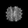
 Map data
 Sample
 Keywords
 Function and homology information
 Homo sapiens (human)
 Authors
 United States, 4 items
  Citation
 Structure visualization
 Downloads & links
 emd_43045.png
 http://ftp.pdbj.org/pub/emdb/structures/EMD-43045
 Z (Sec.)
 Y (Row.)
 X (Col.)


 Sample components
 Processing
 Electron microscopy
 FIELD EMISSION GUN

