Lederhausens Center for CF Research at University Gothenburg
Sweden
Center for Innovative Medicine (KI)
Sweden
Cystic Fibrosis Foundation
14X0
United States
Swedish CF Foundation
Sweden
Cystic Fibrosis Foundation
077X0
United States
Swedish Research Council
Sweden
The Knut and Alice Wallenberg Foundation
Sweden
The Swedish Cancer Foundation
Sweden
Sahlgrenska University Hospital (ALF)
Sweden
IngaBritt and Arne Lundberg Foundation
Sweden
National Institute of Allergy and Infectious Diseases
U01AI095473
Sweden
Wilhelm and Martina Lundgrens Foundation
Sweden
Erica Lederhausens Foundation
Sweden
The Swedish Foundation for Strategic Research
Sweden
Citation
Journal: J Biol Chem / Year: 2018 Title: Granule-stored MUC5B mucins are packed by the non-covalent formation of N-terminal head-to-head tetramers. Authors: Sergio Trillo-Muyo / Harriet E Nilsson / Christian V Recktenwald / Anna Ermund / Caroline Ridley / Lauren N Meiss / Andrea Bähr / Nikolai Klymiuk / Jeffrey J Wine / Philip J B Koeck / David ...Authors: Sergio Trillo-Muyo / Harriet E Nilsson / Christian V Recktenwald / Anna Ermund / Caroline Ridley / Lauren N Meiss / Andrea Bähr / Nikolai Klymiuk / Jeffrey J Wine / Philip J B Koeck / David J Thornton / Hans Hebert / Gunnar C Hansson / Abstract: Most MUC5B mucin polymers in the upper airways of humans and pigs are produced by submucosal glands. MUC5B forms N-terminal covalent dimers that are further packed into larger assemblies because of ...Most MUC5B mucin polymers in the upper airways of humans and pigs are produced by submucosal glands. MUC5B forms N-terminal covalent dimers that are further packed into larger assemblies because of low pH and high Ca in the secretory granule of the mucin-producing cell. We purified the recombinant MUC5B N-terminal covalent dimer and used single-particle electron microscopy to study its structure under intracellular conditions. We found that, at intragranular pH, the dimeric MUC5B organized into head-to-head noncovalent tetramers where the von Willebrand D1-D2 domains hooked into each other. These N-terminal tetramers further formed long linear complexes from which, we suggest, the mucin domains and their C termini project radially outwards. Using conventional and video microscopy, we observed that, upon secretion into the submucosal gland ducts, a flow of bicarbonate-rich fluid passes the mucin-secreting cells. We suggest that this unfolds and pulls out the MUC5B assemblies into long linear threads. These further assemble into thicker mucin bundles in the glandular ducts before emerging at the gland duct opening. We conclude that the combination of intracellular packing of the MUC5B mucin and the submucosal gland morphology creates an efficient machine for producing linear mucin bundles.
History
Deposition
Feb 13, 2018
-
Header (metadata) release
Feb 21, 2018
-
Map release
Mar 20, 2019
-
Update
Oct 2, 2019
-
Current status
Oct 2, 2019
Processing site: PDBe / Status: Released
-
Structure visualization
Movie
Surface view with section colored by density value
Supramolecule #1: MUC5B N-terminal oligomeric state
Supramolecule
Name: MUC5B N-terminal oligomeric state / type: complex / ID: 1 / Parent: 0 Details: The MUC5B N-terminal covalent dimers form non-covalent tetramers at low pH, high Ca2+ in the secretory vesicles of the mucus secreting cells
Source (natural)
Organism: Homo sapiens (human) / Strain: Human / Organ: submucosal gland / Tissue: gland
Number selected: 4456 Details: Negative monitor contrast fascilated particle picking
CTF correction
Software - Name: EMAN2 (ver. 2.12) Software - details: EMAN2 e2boxer.py was used to automatically select particle images Details: For low resolution data EMAN2 uses a 1D structure factor during the CTF procedure
Startup model
Type of model: OTHER Details: The initial model was based on a subset of class averages representing different views.
Final reconstruction
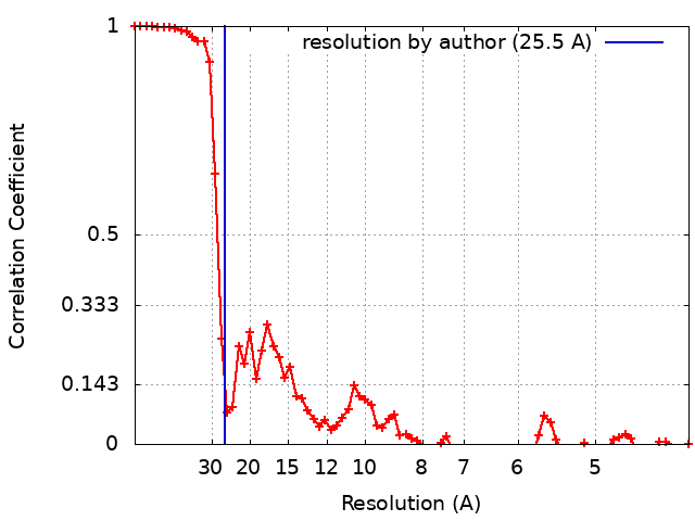
Applied symmetry - Point group: D2 (2x2 fold dihedral) / Algorithm: FOURIER SPACE / Resolution.type: BY AUTHOR / Resolution: 25.5 Å / Resolution method: FSC 0.143 CUT-OFF / Software - Name: EMAN2 (ver. 2.12) / Number images used: 4056
In the structure databanks used in Yorodumi, some data are registered as the other names, "COVID-19 virus" and "2019-nCoV". Here are the details of the virus and the list of structure data.
Jan 31, 2019. EMDB accession codes are about to change! (news from PDBe EMDB page)
EMDB accession codes are about to change! (news from PDBe EMDB page)
The allocation of 4 digits for EMDB accession codes will soon come to an end. Whilst these codes will remain in use, new EMDB accession codes will include an additional digit and will expand incrementally as the available range of codes is exhausted. The current 4-digit format prefixed with “EMD-” (i.e. EMD-XXXX) will advance to a 5-digit format (i.e. EMD-XXXXX), and so on. It is currently estimated that the 4-digit codes will be depleted around Spring 2019, at which point the 5-digit format will come into force.
The EM Navigator/Yorodumi systems omit the EMD- prefix.
Related info.:Q: What is EMD? / ID/Accession-code notation in Yorodumi/EM Navigator
Yorodumi is a browser for structure data from EMDB, PDB, SASBDB, etc.
This page is also the successor to EM Navigator detail page, and also detail information page/front-end page for Omokage search.
The word "yorodu" (or yorozu) is an old Japanese word meaning "ten thousand". "mi" (miru) is to see.
Related info.:EMDB / PDB / SASBDB / Comparison of 3 databanks / Yorodumi Search / Aug 31, 2016. New EM Navigator & Yorodumi / Yorodumi Papers / Jmol/JSmol / Function and homology information / Changes in new EM Navigator and Yorodumi
 Movie
Movie Controller


 Open data
 Basic information
 Map data
 Sample
 Homo sapiens (human)
 Authors
 Sweden,
  United States, 16 items
  Citation

 Structure visualization
 Movie viewer
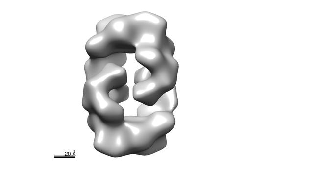
 Downloads & links
 emd_4296.png
 http://ftp.pdbj.org/pub/emdb/structures/EMD-4296


 Z (Sec.)
 Y (Row.)
 X (Col.)


 Sample components
 Cricetinae gen. sp. (mammal) / Recombinant strain: ovary / Recombinant cell: CHO-K1 cells
 Processing
 Electron microscopy
 FIELD EMISSION GUN