 Movie
Movie Controller

[English] 日本語
 Yorodumi
- EMDB-42113: Tomograms of the apical end of Toxoplasma Gondii with Rhoptry Dis... -
+ Open data

- Basic information
| Entry |  | |||||||||||||||
|---|---|---|---|---|---|---|---|---|---|---|---|---|---|---|---|---|
| Title | Tomograms of the apical end of Toxoplasma Gondii with Rhoptry Discharge Factor 3 (RDF3) conditionally knocked down | |||||||||||||||
 Map data Map data | Toxoplasma gondii with Rhoptry Discharge Factor 3 (RDF3) conditionally knocked down | |||||||||||||||
 Sample Sample |
| |||||||||||||||
 Keywords Keywords | Rhoptry / Parasite / Conoid / CELL INVASION | |||||||||||||||
| Biological species |  | |||||||||||||||
| Method | electron tomography / cryo EM | |||||||||||||||
 Authors Authors | Martinez M / Ben Chaabene R / Chang Y-W / Soldati-Favre D | |||||||||||||||
| Funding support |  United States, United States,  Switzerland, European Union, 4 items Switzerland, European Union, 4 items
| |||||||||||||||
 Citation Citation | Journal: PLoS Biol / Year: 2024 Title: Toxoplasma gondii rhoptry discharge factor 3 is essential for invasion and microtubule-associated vesicle biogenesis. Authors: Rouaa Ben Chaabene / Matthew Martinez / Alessandro Bonavoglia / Bohumil Maco / Yi-Wei Chang / Gaëlle Lentini / Dominique Soldati-Favre / Abstract: Rhoptries are specialized secretory organelles conserved across the Apicomplexa phylum, essential for host cell invasion and critical for subverting of host cellular and immune functions. They ...Rhoptries are specialized secretory organelles conserved across the Apicomplexa phylum, essential for host cell invasion and critical for subverting of host cellular and immune functions. They contain proteins and membranous materials injected directly into the host cells, participating in parasitophorous vacuole formation. Toxoplasma gondii tachyzoites harbor 8 to 12 rhoptries, 2 of which are docked to an apical vesicle (AV), a central element associated with a rhoptry secretory apparatus prior to injection into the host cell. This parasite is also equipped with 5 to 6 microtubule-associated vesicles, presumably serving as AV replenishment for iterative rhoptry discharge. Here, we characterized a rhoptry protein, rhoptry discharge factor 3 (RDF3), crucial for rhoptry discharge and invasion. RDF3 enters the secretory pathway, localizing near the AV and associated with the rhoptry bulb. Upon invasion, RDF3 dynamically delocalizes, suggesting a critical role at the time of rhoptry discharge. Cryo-electron tomography analysis of RDF3-depleted parasites reveals irregularity in microtubule-associated vesicles morphology, presumably impacting on their preparedness to function as an AV. Our findings suggest that RDF3 is priming the microtubule-associated vesicles for rhoptry discharge by a mechanism distinct from the rhoptry secretory apparatus contribution. | |||||||||||||||
| History |
|
- Structure visualization
| Supplemental images |
|---|

- Downloads & links
-EMDB archive
| Map data | emd_42113.map.gz | 1.3 GB |  EMDB map data format EMDB map data format | |
|---|---|---|---|---|
| Header (meta data) | emd-42113-v30.xmlemd-42113.xml | 14.3 KB 14.3 KB | Display Display | EMDB header |
| Images |  emd_42113.png emd_42113.png | 122.7 KB | ||
| Filedesc metadata | emd-42113.cif.gz | 4.4 KB | ||
| Others | emd_42113_additional_1.map.gz | 1.2 GB | ||
| Archive directory |  http://ftp.pdbj.org/pub/emdb/structures/EMD-42113ftp://ftp.pdbj.org/pub/emdb/structures/EMD-42113 http://ftp.pdbj.org/pub/emdb/structures/EMD-42113ftp://ftp.pdbj.org/pub/emdb/structures/EMD-42113 | HTTPS FTP |
-Related structure data
-Links
| EMDB pages | EMDB (EBI/PDBe) / EMDataResource |
|---|
-Map
| File | Download / File: emd_42113.map.gz / Format: CCP4 / Size: 2.2 GB / Type: IMAGE STORED AS SIGNED INTEGER (2 BYTES) | ||||||||||||||||||||||||||||||||
|---|---|---|---|---|---|---|---|---|---|---|---|---|---|---|---|---|---|---|---|---|---|---|---|---|---|---|---|---|---|---|---|---|---|
| Annotation | Toxoplasma gondii with Rhoptry Discharge Factor 3 (RDF3) conditionally knocked down | ||||||||||||||||||||||||||||||||
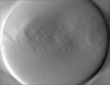
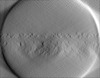
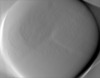
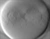
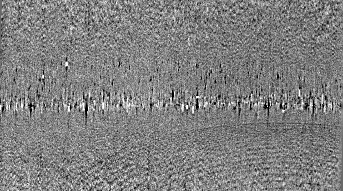
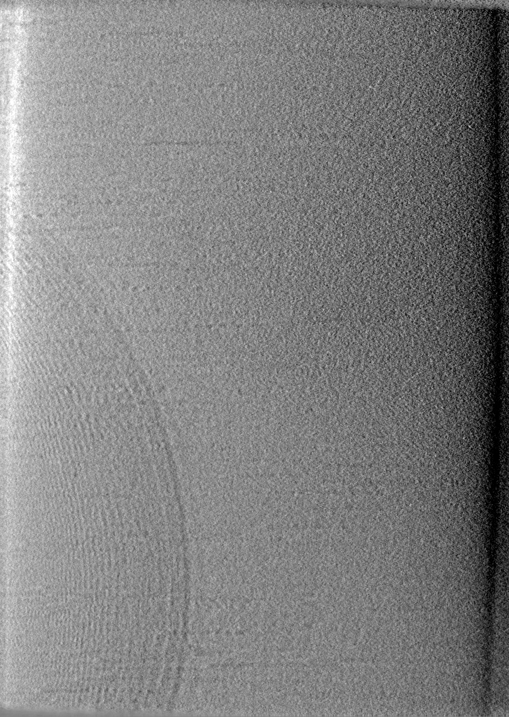
| Projections & slices | Image control
Images are generated by Spider. generated in cubic-lattice coordinate | ||||||||||||||||||||||||||||||||
| Voxel size | X=Y=Z: 10.6 Å | ||||||||||||||||||||||||||||||||
| Density |
| ||||||||||||||||||||||||||||||||
| Symmetry | Space group: 1 | ||||||||||||||||||||||||||||||||
| Details | EMDB XML:
|
 Z (Sec.)
 Y (Row.)
 X (Col.)


-Supplemental data
-Additional map: Toxoplasma gondii with Rhoptry Discharge Factor 3 (RDF3)...
| File | emd_42113_additional_1.map | ||||||||||||
|---|---|---|---|---|---|---|---|---|---|---|---|---|---|
| Annotation | Toxoplasma gondii with Rhoptry Discharge Factor 3 (RDF3) conditionally knocked down | ||||||||||||
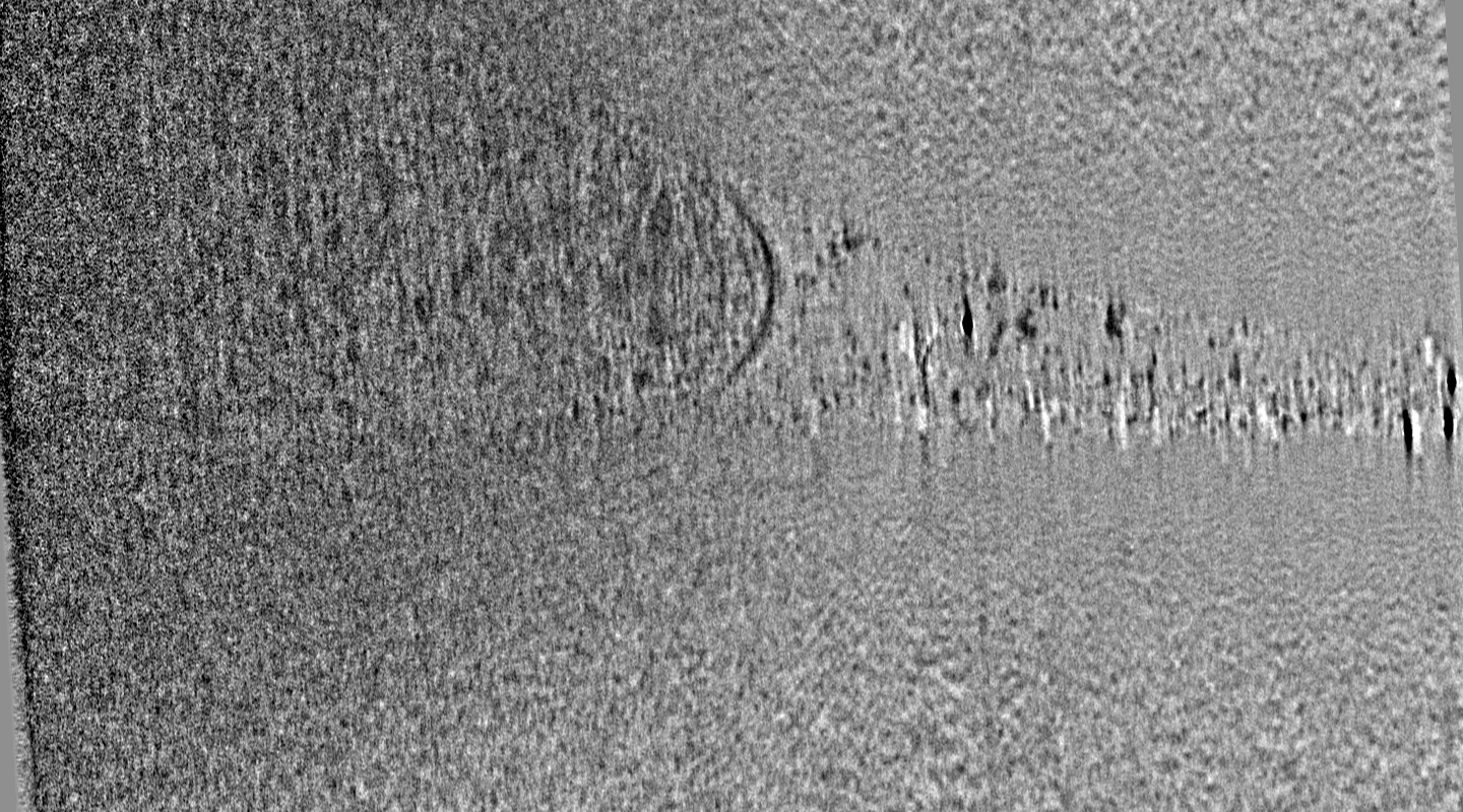
| Projections & Slices |
| ||||||||||||
| Density Histograms |


- Sample components
-Entire : Toxoplasma Gondii tachyzoite apical complex with Rhoptry Discharg...
| Entire | Name: Toxoplasma Gondii tachyzoite apical complex with Rhoptry Discharge Factor 3 (RDF3) conditionally knocked down |
|---|---|
| Components |
|
-Supramolecule #1: Toxoplasma Gondii tachyzoite apical complex with Rhoptry Discharg...
| Supramolecule | Name: Toxoplasma Gondii tachyzoite apical complex with Rhoptry Discharge Factor 3 (RDF3) conditionally knocked down type: cell / ID: 1 / Parent: 0 |
|---|---|
| Source (natural) | Organism: |
-Experimental details
-Structure determination
| Method | cryo EM |
|---|---|
 Processing Processing | electron tomography |
| Aggregation state | cell |
-Sample preparation
| Buffer | pH: 7.4 Details: DMEM supplemented with 5% fetal bovine serum and 2 mM glutamine |
|---|---|
| Grid | Model: Quantifoil R2/2 / Material: COPPER / Mesh: 200 / Support film - Material: CARBON / Support film - topology: HOLEY |
| Vitrification | Cryogen name: ETHANE-PROPANE / Chamber humidity: 95 % / Chamber temperature: 310 K / Instrument: LEICA EM GP / Details: Front blot for 4 seconds before plunging. |
| Details | Egressed tachyzoites from a culture of confluent HFF cells. RDF3 knockdown induced by treatment with 50 nM rapamycin for 48 hours prior to egress. |
| Sectioning | Other: NO SECTIONING |
| Fiducial marker | Manufacturer: Ted Pella, Inc / Diameter: 10 nm |
- Electron microscopy
| Microscope | FEI TITAN KRIOS |
|---|---|
| Specialist optics | Phase plate: VOLTA PHASE PLATE / Energy filter - Name: GIF Bioquantum / Energy filter - Slit width: 20 eV |
| Software | Name: SerialEM (ver. 3.8) |
| Image recording | Film or detector model: GATAN K3 (6k x 4k) / Average exposure time: 0.4 sec. / Average electron dose: 2.3 e/Å2 |
| Electron beam | Acceleration voltage: 300 kV / Electron source:  FIELD EMISSION GUN FIELD EMISSION GUN |
| Electron optics | C2 aperture diameter: 100.0 µm / Illumination mode: FLOOD BEAM / Imaging mode: BRIGHT FIELD / Nominal defocus max: 4.0 µm / Nominal defocus min: 1.5 µm / Nominal magnification: 33000 |
| Sample stage | Specimen holder model: FEI TITAN KRIOS AUTOGRID HOLDER / Cooling holder cryogen: NITROGEN |
| Experimental equipment |  Model: Titan Krios / Image courtesy: FEI Company |
-Image processing
| Details | Raw frames were gain normalized in serialEM and motion corrected using the alignframes command in IMOD |
|---|---|
| Final reconstruction | Algorithm: BACK PROJECTION / Software - Name: IMOD (ver. 4.11.5) / Number images used: 61 |
