ribonucleotide binding / DNA primase AEP / DNA replication initiation / DNA/RNA hybrid binding / Inhibition of replication initiation of damaged DNA by RB1/E2F1 / Telomere C-strand synthesis initiation / alpha DNA polymerase:primase complex / regulation of type I interferon production / Polymerase switching / Processive synthesis on the lagging strand ...ribonucleotide binding / DNA primase AEP / DNA replication initiation / DNA/RNA hybrid binding / Inhibition of replication initiation of damaged DNA by RB1/E2F1 / Telomere C-strand synthesis initiation / alpha DNA polymerase:primase complex / regulation of type I interferon production / Polymerase switching / Processive synthesis on the lagging strand / Removal of the Flap Intermediate / lagging strand elongation / DNA replication, synthesis of primer / mitotic DNA replication initiation / Polymerase switching on the C-strand of the telomere / DNA strand elongation involved in DNA replication / G1/S-Specific Transcription / leading strand elongation / DNA synthesis involved in DNA repair / DNA replication origin binding / Activation of the pre-replicative complex / DNA replication initiation / Defective pyroptosis / double-strand break repair via nonhomologous end joining / nuclear matrix / protein import into nucleus / DNA-directed RNA polymerase activity / nuclear envelope / single-stranded DNA binding / 4 iron, 4 sulfur cluster binding / DNA-directed DNA polymerase / DNA-directed DNA polymerase activity / DNA replication / ciliary basal body / nucleotide binding / DNA repair / chromatin binding / protein kinase binding / chromatin / nucleolus / magnesium ion binding / DNA binding / zinc ion binding / nucleoplasm / membrane / metal ion binding / nucleus / cytosol Similarity search - Function
DNA polymerase alpha, subunit B, N-terminal domain superfamily / : / DNA polymerase alpha subunit B, OB domain / DNA polymerase alpha subunit B N-terminal / DNA polymerase alpha, subunit B, N-terminal / DNA polymerase alpha, subunit B / : / Eukaryotic and archaeal DNA primase, large subunit N-terminal domain / DNA primase, small subunit, eukaryotic/archaeal / DNA primase large subunit, eukaryotic/archaeal ...DNA polymerase alpha, subunit B, N-terminal domain superfamily / : / DNA polymerase alpha subunit B, OB domain / DNA polymerase alpha subunit B N-terminal / DNA polymerase alpha, subunit B, N-terminal / DNA polymerase alpha, subunit B / : / Eukaryotic and archaeal DNA primase, large subunit N-terminal domain / DNA primase, small subunit, eukaryotic/archaeal / DNA primase large subunit, eukaryotic/archaeal / DNA primase, large subunit, eukaryotic / DNA primase, small subunit / DNA primase small subunit / DNA polymerase alpha catalytic subunit, N-terminal domain / DNA polymerase alpha, zinc finger domain superfamily / Eukaryotic and archaeal DNA primase, large subunit C-terminal domain / DNA Polymerase alpha zinc finger / DNA polymerase alpha subunit p180 N terminal / Zinc finger, DNA-directed DNA polymerase, family B, alpha / DNA polymerase alpha catalytic subunit, catalytic domain / DNA polymerase alpha/delta/epsilon, subunit B / DNA polymerase alpha/epsilon subunit B / DNA polymerase family B, thumb domain / DNA-directed DNA polymerase, family B, multifunctional domain / DNA-directed DNA polymerase, family B, conserved site / DNA polymerase family B signature. / DNA polymerase family B / DNA polymerase family B, exonuclease domain / DNA-directed DNA polymerase, family B, exonuclease domain / DNA polymerase, palm domain superfamily / DNA polymerase type-B family / DNA-directed DNA polymerase, family B / Ribonuclease H superfamily / Ribonuclease H-like superfamily / DNA/RNA polymerase superfamily Similarity search - Domain/homology
DNA polymerase alpha catalytic subunit / DNA primase small subunit / DNA primase large subunit / DNA polymerase alpha subunit B Similarity search - Component
Biological species
Homo sapiens (human)
Method
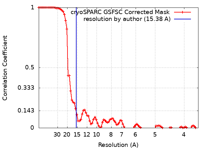
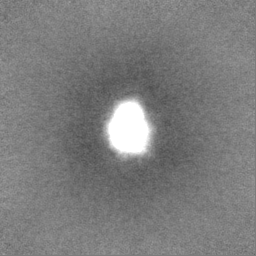
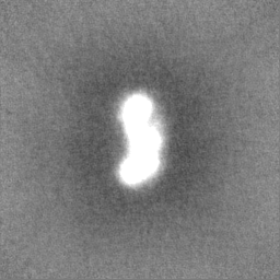
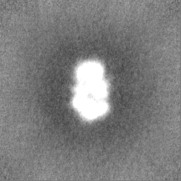
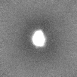
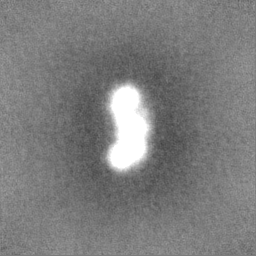
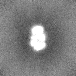
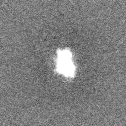
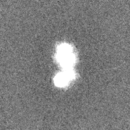
single particle reconstruction / negative staining / Resolution: 15.38 Å
National Institutes of Health/National Institute of General Medical Sciences (NIH/NIGMS)
2T32GM008320-31
United States
National Institutes of Health/National Institute of General Medical Sciences (NIH/NIGMS)
R35GM118089
United States
National Institutes of Health/National Institute of General Medical Sciences (NIH/NIGMS)
R35GM136401
United States
Citation
Journal: J Mol Biol / Year: 2023 Title: Flexibility and Distributive Synthesis Regulate RNA Priming and Handoff in Human DNA Polymerase α-Primase. Authors: John J Cordoba / Elwood A Mullins / Lauren E Salay / Brandt F Eichman / Walter J Chazin / Abstract: DNA replication in eukaryotes relies on the synthesis of a ∼30-nucleotide RNA/DNA primer strand through the dual action of the heterotetrameric polymerase α-primase (pol-prim) enzyme. Synthesis of ...DNA replication in eukaryotes relies on the synthesis of a ∼30-nucleotide RNA/DNA primer strand through the dual action of the heterotetrameric polymerase α-primase (pol-prim) enzyme. Synthesis of the 7-10-nucleotide RNA primer is regulated by the C-terminal domain of the primase regulatory subunit (PRIM2C) and is followed by intramolecular handoff of the primer to pol α for extension by ∼20 nucleotides of DNA. Here, we provide evidence that RNA primer synthesis is governed by a combination of the high affinity and flexible linkage of the PRIM2C domain and the surprisingly low affinity of the primase catalytic domain (PRIM1) for substrate. Using a combination of small angle X-ray scattering and electron microscopy, we found significant variability in the organization of PRIM2C and PRIM1 in the absence and presence of substrate, and that the population of structures with both PRIM2C and PRIM1 in a configuration aligned for synthesis is low. Crosslinking was used to visualize the orientation of PRIM2C and PRIM1 when engaged by substrate as observed by electron microscopy. Microscale thermophoresis was used to measure substrate affinities for a series of pol-prim constructs, which showed that the PRIM1 catalytic domain does not bind the template or emergent RNA-primed templates with appreciable affinity. Together, these findings support a model of RNA primer synthesis in which generation of the nascent RNA strand and handoff of the RNA-primed template from primase to polymerase α is mediated by the high degree of inter-domain flexibility of pol-prim, the ready dissociation of PRIM1 from its substrate, and the much higher affinity of the POLA1cat domain of polymerase α for full-length RNA-primed templates.
Name: RNA priming substrate - template / type: dna / ID: 6 / Classification: DNA
Source (natural)
Organism: Homo sapiens (human)
Sequence
String:
GTATGTATGT CAGTATCCTG TATGTATGA
-
Experimental details
-
Structure determination
Method
negative staining
Processing
single particle reconstruction
Aggregation state
particle
-
Sample preparation
Concentration
0.009 mg/mL
Buffer
pH: 7.5 Component:
Concentration
Formula
Name
150.0 mM
NaCl
Sodium chloride
20.0 mM
C8H18N2O4S
HEPES
5.0 mM
MgCl2
Magnesium chloride
1.0 mM
C9H15O6P
TCEP
Staining
Type: NEGATIVE / Material: Uranyl Formate Details: 2.5 uL sample was applied to grid for 1 minute, then blotted with filter paper, washed for 5 seconds with DI water, blotted, washed for 5 seconds in stain, blotted, then stained for 90 seconds.
-
Electron microscopy
Microscope
FEI TECNAI 20
Image recording
Film or detector model: GATAN ULTRASCAN 4000 (4k x 4k) / Digitization - Dimensions - Width: 4096 pixel / Digitization - Dimensions - Height: 4096 pixel / Number grids imaged: 1 / Number real images: 1083 / Average electron dose: 20.0 e/Å2
Electron beam
Acceleration voltage: 200 kV / Electron source: FIELD EMISSION GUN
In the structure databanks used in Yorodumi, some data are registered as the other names, "COVID-19 virus" and "2019-nCoV". Here are the details of the virus and the list of structure data.
Jan 31, 2019. EMDB accession codes are about to change! (news from PDBe EMDB page)
EMDB accession codes are about to change! (news from PDBe EMDB page)
The allocation of 4 digits for EMDB accession codes will soon come to an end. Whilst these codes will remain in use, new EMDB accession codes will include an additional digit and will expand incrementally as the available range of codes is exhausted. The current 4-digit format prefixed with “EMD-” (i.e. EMD-XXXX) will advance to a 5-digit format (i.e. EMD-XXXXX), and so on. It is currently estimated that the 4-digit codes will be depleted around Spring 2019, at which point the 5-digit format will come into force.
The EM Navigator/Yorodumi systems omit the EMD- prefix.
Related info.:Q: What is EMD? / ID/Accession-code notation in Yorodumi/EM Navigator
Yorodumi is a browser for structure data from EMDB, PDB, SASBDB, etc.
This page is also the successor to EM Navigator detail page, and also detail information page/front-end page for Omokage search.
The word "yorodu" (or yorozu) is an old Japanese word meaning "ten thousand". "mi" (miru) is to see.
Related info.:EMDB / PDB / SASBDB / Comparison of 3 databanks / Yorodumi Search / Aug 31, 2016. New EM Navigator & Yorodumi / Yorodumi Papers / Jmol/JSmol / Function and homology information / Changes in new EM Navigator and Yorodumi
 Movie
Movie Controller

 Yorodumi
 Open data
 Basic information
 Map data
 Sample
 Keywords
 Function and homology information
 Homo sapiens (human)
 Authors
 United States, 3 items
  Citation
 Structure visualization
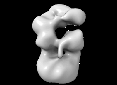
 Downloads & links
 emd_42034.png
 http://ftp.pdbj.org/pub/emdb/structures/EMD-42034


 Z (Sec.)
 Y (Row.)
 X (Col.)


 Sample components
 Processing
 Electron microscopy
 FIELD EMISSION GUN