 Movie
Movie Controller

[English] 日本語
 Yorodumi
- EMDB-42009: KCTD5/Cullin3/Gbeta1gamma2 Complex State D: Reference Map for Com... -
+ Open data

- Basic information
| Entry |  | |||||||||
|---|---|---|---|---|---|---|---|---|---|---|
| Title | KCTD5/Cullin3/Gbeta1gamma2 Complex State D: Reference Map for Composite Map EMD-42008 | |||||||||
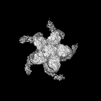
 Map data Map data | KCTD5:Cullin3:GBetaGamma StateD Reference Map: Full Map | |||||||||
 Sample Sample |
| |||||||||
 Keywords Keywords | cullin family protein / proteasome-mediated ubiquitin-dependent protein catabolic process / complex / ubiquitin-dependent protein catabolic process / CYTOSOLIC PROTEIN | |||||||||
| Biological species |  Homo sapiens (human) Homo sapiens (human) | |||||||||
| Method | single particle reconstruction / cryo EM / Resolution: 3.98 Å | |||||||||
 Authors Authors | Nguyen DM / Narayanan N / Kuntz DA / Prive GG | |||||||||
| Funding support |  Canada, 1 items Canada, 1 items
| |||||||||
 Citation Citation | Journal: Proc Natl Acad Sci U S A / Year: 2024 Title: Structure and dynamics of a pentameric KCTD5/CUL3/Gβγ E3 ubiquitin ligase complex. Authors: Duc Minh Nguyen / Deanna H Rath / Dominic Devost / Darlaine Pétrin / Robert Rizk / Alan X Ji / Naveen Narayanan / Darren Yong / Andrew Zhai / Douglas A Kuntz / Maha U Q Mian / Neil C Pomroy ...Authors: Duc Minh Nguyen / Deanna H Rath / Dominic Devost / Darlaine Pétrin / Robert Rizk / Alan X Ji / Naveen Narayanan / Darren Yong / Andrew Zhai / Douglas A Kuntz / Maha U Q Mian / Neil C Pomroy / Alexander F A Keszei / Samir Benlekbir / Mohammad T Mazhab-Jafari / John L Rubinstein / Terence E Hébert / Gilbert G Privé / Abstract: Heterotrimeric G proteins can be regulated by posttranslational modifications, including ubiquitylation. KCTD5, a pentameric substrate receptor protein consisting of an N-terminal BTB domain and a C- ...Heterotrimeric G proteins can be regulated by posttranslational modifications, including ubiquitylation. KCTD5, a pentameric substrate receptor protein consisting of an N-terminal BTB domain and a C-terminal domain, engages CUL3 to form the central scaffold of a cullin-RING E3 ligase complex (CRL3) that ubiquitylates Gβγ and reduces Gβγ protein levels in cells. The cryo-EM structure of a 5:5:5 KCTD5/CUL3/Gβγ assembly reveals a highly dynamic complex with rotations of over 60° between the KCTD5/CUL3 and KCTD5/Gβγ moieties of the structure. CRL3 engages the E3 ligase ARIH1 to ubiquitylate Gβγ in an E3-E3 superassembly, and extension of the structure to include full-length CUL3 with RBX1 and an ARIH1~ubiquitin conjugate reveals that some conformational states position the ARIH1~ubiquitin thioester bond to within 10 Å of lysine-23 of Gβ and likely represent priming complexes. Most previously described CRL/substrate structures have consisted of monovalent complexes and have involved flexible peptide substrates. The structure of the KCTD5/CUL3/Gβγ complex shows that the oligomerization of a substrate receptor can generate a polyvalent E3 ligase complex and that the internal dynamics of the substrate receptor can position a structured target for ubiquitylation in a CRL3 complex. #1: Journal: Biorxiv / Year: 2023Title: Structure and dynamics of a pentameric KCTD5/Cullin3/G beta gamma E3 ubiquitin ligase complex Authors: Nguyen DM / Rath DH / Devost D / Petrin D / Rizk R / Ji AX / Narayanan N / Yong D / Zhai A / Kuntz DA / Mian MUQ / Pomroy NC / Keszei AFE / Benlekbir S / Mazhab-Jafari MT / Rubinstein JL / Hebert TE / Prive GG | |||||||||
| History |
|
- Structure visualization
| Supplemental images |
|---|

- Downloads & links
-EMDB archive
| Map data | emd_42009.map.gz | 101.5 MB |  EMDB map data format EMDB map data format | |
|---|---|---|---|---|
| Header (meta data) | emd-42009-v30.xmlemd-42009.xml | 15.7 KB 15.7 KB | Display Display | EMDB header |
| FSC (resolution estimation) | emd_42009_fsc.xml | 11.5 KB | Display | FSC data file |
| Images |  emd_42009.png emd_42009.png | 111.7 KB | ||
| Masks | emd_42009_msk_1.map | 129.7 MB | Mask map | |
| Filedesc metadata | emd-42009.cif.gz | 4.6 KB | ||
| Others | emd_42009_half_map_1.map.gzemd_42009_half_map_2.map.gz | 102 MB 102 MB | ||
| Archive directory |  http://ftp.pdbj.org/pub/emdb/structures/EMD-42009ftp://ftp.pdbj.org/pub/emdb/structures/EMD-42009 http://ftp.pdbj.org/pub/emdb/structures/EMD-42009ftp://ftp.pdbj.org/pub/emdb/structures/EMD-42009 | HTTPS FTP |
-Related structure data


-Links
| EMDB pages | EMDB (EBI/PDBe) / EMDataResource |
|---|
-Map
| File | Download / File: emd_42009.map.gz / Format: CCP4 / Size: 129.7 MB / Type: IMAGE STORED AS FLOATING POINT NUMBER (4 BYTES) | ||||||||||||||||||||||||||||||||||||
|---|---|---|---|---|---|---|---|---|---|---|---|---|---|---|---|---|---|---|---|---|---|---|---|---|---|---|---|---|---|---|---|---|---|---|---|---|---|
| Annotation | KCTD5:Cullin3:GBetaGamma StateD Reference Map: Full Map | ||||||||||||||||||||||||||||||||||||
| Projections & slices | Image control
Images are generated by Spider. | ||||||||||||||||||||||||||||||||||||
| Voxel size | X=Y=Z: 1.03 Å | ||||||||||||||||||||||||||||||||||||
| Density |
| ||||||||||||||||||||||||||||||||||||
| Symmetry | Space group: 1 | ||||||||||||||||||||||||||||||||||||
| Details | EMDB XML:
|
 Z (Sec.)
 Y (Row.)
 X (Col.)


-Supplemental data
-Mask #1
| File | emd_42009_msk_1.map | ||||||||||||
|---|---|---|---|---|---|---|---|---|---|---|---|---|---|
| Projections & Slices |
| ||||||||||||
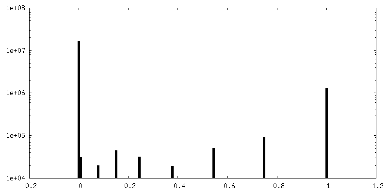
| Density Histograms |


-Half map: KCTD5:Cullin3:GBetaGamma StateD Reference Map: Half Map 1
| File | emd_42009_half_map_1.map | ||||||||||||
|---|---|---|---|---|---|---|---|---|---|---|---|---|---|
| Annotation | KCTD5:Cullin3:GBetaGamma StateD Reference Map: Half Map 1 | ||||||||||||
| Projections & Slices |
| ||||||||||||
| Density Histograms |


-Half map: KCTD5:Cullin3:GBetaGamma StateD Reference Map: Half Map 2
| File | emd_42009_half_map_2.map | ||||||||||||
|---|---|---|---|---|---|---|---|---|---|---|---|---|---|
| Annotation | KCTD5:Cullin3:GBetaGamma StateD Reference Map: Half Map 2 | ||||||||||||
| Projections & Slices |
| ||||||||||||
| Density Histograms |


- Sample components
-Entire : Complex of substrate adaptor KCTD5 with Cullin-3 and substrate Gb...
| Entire | Name: Complex of substrate adaptor KCTD5 with Cullin-3 and substrate Gbeta-gamma |
|---|---|
| Components |
|
-Supramolecule #1: Complex of substrate adaptor KCTD5 with Cullin-3 and substrate Gb...
| Supramolecule | Name: Complex of substrate adaptor KCTD5 with Cullin-3 and substrate Gbeta-gamma type: complex / ID: 1 / Parent: 0 |
|---|---|
| Source (natural) | Organism: Homo sapiens (human) |
-Experimental details
-Structure determination
| Method | cryo EM |
|---|---|
 Processing Processing | single particle reconstruction |
| Aggregation state | particle |
-Sample preparation
| Buffer | pH: 7.5 |
|---|---|
| Vitrification | Cryogen name: ETHANE / Instrument: FEI VITROBOT MARK IV |
- Electron microscopy
| Microscope | FEI TITAN KRIOS |
|---|---|
| Image recording | Film or detector model: FEI FALCON IV (4k x 4k) / Number real images: 7464 / Average electron dose: 49.35 e/Å2 |
| Electron beam | Acceleration voltage: 300 kV / Electron source:  FIELD EMISSION GUN FIELD EMISSION GUN |
| Electron optics | Illumination mode: FLOOD BEAM / Imaging mode: BRIGHT FIELD / Nominal defocus max: 1.5 µm / Nominal defocus min: 0.25 µm / Nominal magnification: 75000 |
| Experimental equipment |  Model: Titan Krios / Image courtesy: FEI Company |
+Image processing

-Atomic model buiding 1
| Refinement | Space: REAL / Protocol: FLEXIBLE FIT |
|---|
