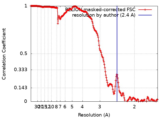
- EMDB-41611: PHF1-Phosphomimetic Tau Filaments (Full-length, Cofactor-Free 0N4... -
+
Open data
ID or keywords:
Loading...
-
Basic information
Entry
Database: EMDB / ID: EMD-41611
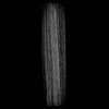
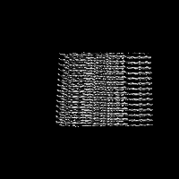
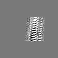
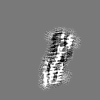
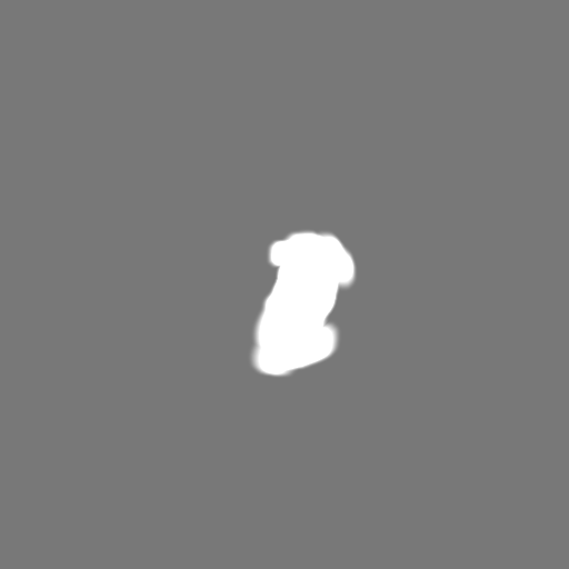
Title
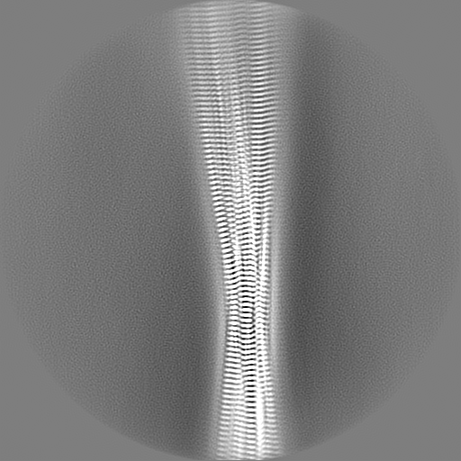
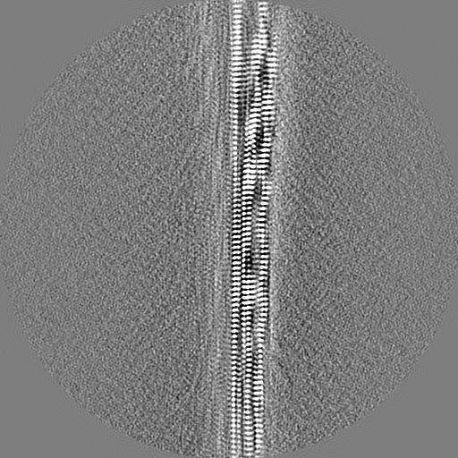
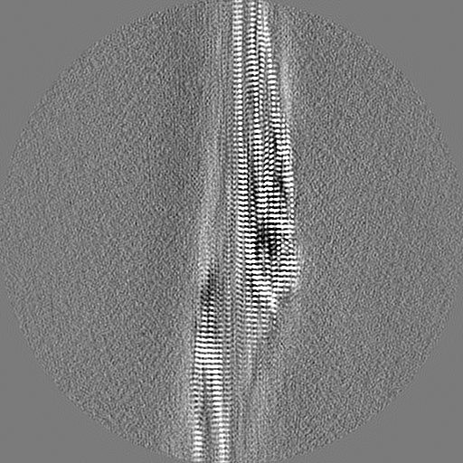
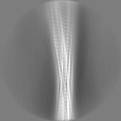
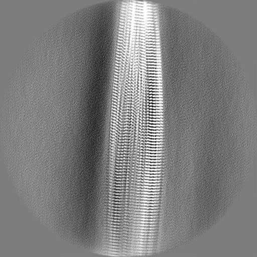
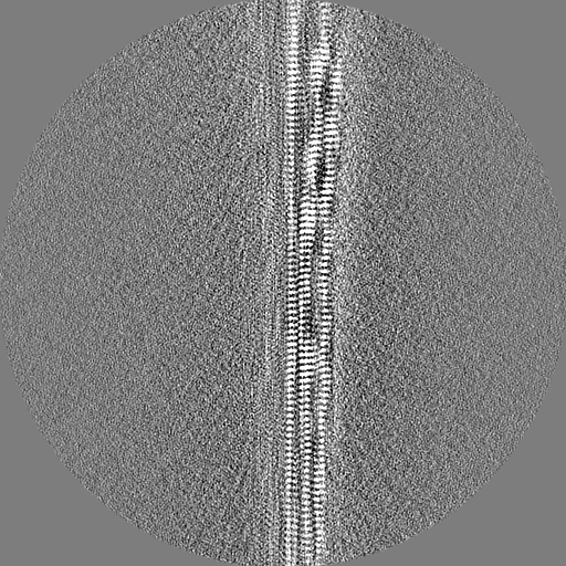
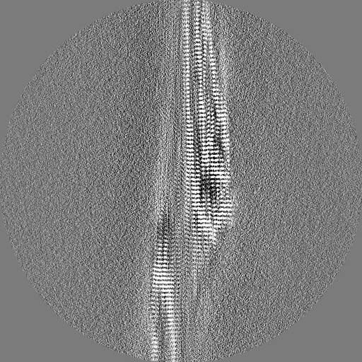
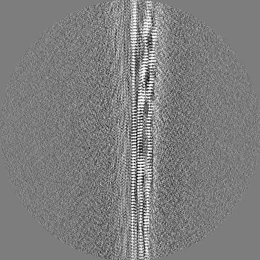
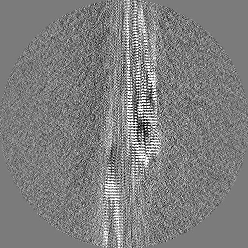
PHF1-Phosphomimetic Tau Filaments (Full-length, Cofactor-Free 0N4R Tau S396E, S400E, T403E, S404E)
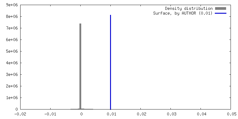
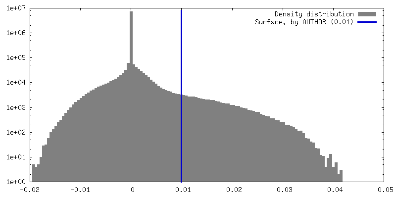
Map data
Sample
Complex: PHF1-Phosphomimetic 0N4R Tau Fibrils
Protein or peptide: Microtubule-associated protein tau
Keywords
Tau / Amyloid Fibril / Phosphomimetic / PROTEIN FIBRIL
Function / homology
Function and homology information
plus-end-directed organelle transport along microtubule / histone-dependent DNA binding / negative regulation of protein localization to mitochondrion / neurofibrillary tangle / microtubule lateral binding / axonal transport / tubulin complex / positive regulation of protein localization to synapse / phosphatidylinositol bisphosphate binding / generation of neurons ...plus-end-directed organelle transport along microtubule / histone-dependent DNA binding / negative regulation of protein localization to mitochondrion / neurofibrillary tangle / microtubule lateral binding / axonal transport / tubulin complex / positive regulation of protein localization to synapse / phosphatidylinositol bisphosphate binding / generation of neurons / rRNA metabolic process / axonal transport of mitochondrion / regulation of mitochondrial fission / axon development / regulation of chromosome organization / regulation of microtubule-based movement / central nervous system neuron development / intracellular distribution of mitochondria / minor groove of adenine-thymine-rich DNA binding / lipoprotein particle binding / microtubule polymerization / negative regulation of mitochondrial membrane potential / regulation of microtubule polymerization / dynactin binding / apolipoprotein binding / main axon / protein polymerization / axolemma / glial cell projection / Caspase-mediated cleavage of cytoskeletal proteins / regulation of microtubule polymerization or depolymerization / negative regulation of mitochondrial fission / neurofibrillary tangle assembly / positive regulation of axon extension / regulation of cellular response to heat / synapse assembly / Activation of AMPK downstream of NMDARs / regulation of long-term synaptic depression / positive regulation of superoxide anion generation / positive regulation of protein localization / cellular response to brain-derived neurotrophic factor stimulus / supramolecular fiber organization / cytoplasmic microtubule organization / regulation of calcium-mediated signaling / somatodendritic compartment / axon cytoplasm / positive regulation of microtubule polymerization / astrocyte activation / phosphatidylinositol binding / enzyme inhibitor activity / nuclear periphery / stress granule assembly / protein phosphatase 2A binding / regulation of microtubule cytoskeleton organization / cellular response to reactive oxygen species / Hsp90 protein binding / microglial cell activation / cellular response to nerve growth factor stimulus / synapse organization / PKR-mediated signaling / regulation of synaptic plasticity / protein homooligomerization / response to lead ion / SH3 domain binding / microtubule cytoskeleton organization / memory / regulation of autophagy / cytoplasmic ribonucleoprotein granule / neuron projection development / cell-cell signaling / single-stranded DNA binding / protein-folding chaperone binding / cellular response to heat / microtubule cytoskeleton / growth cone / actin binding / cell body / double-stranded DNA binding / protein-macromolecule adaptor activity / microtubule binding / sequence-specific DNA binding / dendritic spine / amyloid fibril formation / microtubule / learning or memory / neuron projection / membrane raft / axon / negative regulation of gene expression / neuronal cell body / DNA damage response / dendrite / protein kinase binding / enzyme binding / mitochondrion / DNA binding / RNA binding / extracellular region / identical protein binding / nucleus Similarity search - Function
Microtubule-associated protein Tau / Microtubule associated protein, tubulin-binding repeat / Tau and MAP protein, tubulin-binding repeat / Tau and MAP proteins tubulin-binding repeat signature. / Tau and MAP proteins tubulin-binding repeat profile. / : Similarity search - Domain/homology
National Institutes of Health/National Institute on Aging (NIH/NIA)
AG059661
United States
National Institutes of Health/National Institute on Aging (NIH/NIA)
AG069418
United States
National Institutes of Health/National Institute of General Medical Sciences (NIH/NIGMS)
GM132079
United States
Citation
Journal: Proc Natl Acad Sci U S A / Year: 2024 Title: Structures of AT8 and PHF1 phosphomimetic tau: Insights into the posttranslational modification code of tau aggregation. Authors: Nadia El Mammeri / Aurelio J Dregni / Pu Duan / Mei Hong / Abstract: The microtubule-associated protein tau aggregates into amyloid fibrils in Alzheimer's disease and other neurodegenerative diseases. In these tauopathies, tau is hyperphosphorylated, suggesting that ...The microtubule-associated protein tau aggregates into amyloid fibrils in Alzheimer's disease and other neurodegenerative diseases. In these tauopathies, tau is hyperphosphorylated, suggesting that this posttranslational modification (PTM) may induce tau aggregation. Tau is also phosphorylated in normal developing brains. To investigate how tau phosphorylation induces amyloid fibrils, here we report the atomic structures of two phosphomimetic full-length tau fibrils assembled without anionic cofactors. We mutated key Ser and Thr residues to Glu in two regions of the protein. One construct contains three Glu mutations at the epitope of the anti-phospho-tau antibody AT8 (AT8-3E tau), whereas the other construct contains four Glu mutations at the epitope of the antibody PHF1 (PHF1-4E tau). Solid-state NMR data show that both phosphomimetic tau mutants form homogeneous fibrils with a single set of chemical shifts. The AT8-3E tau rigid core extends from the R3 repeat to the C terminus, whereas the PHF1-4E tau rigid core spans R2, R3, and R4 repeats. Cryoelectron microscopy data show that AT8-3E tau forms a triangular multi-layered core, whereas PHF1-4E tau forms a triple-stranded core. Interestingly, a construct combining all seven Glu mutations exhibits the same conformation as PHF1-4E tau. Scalar-coupled NMR data additionally reveal the dynamics and shape of the fuzzy coat surrounding the rigid cores. These results demonstrate that specific PTMs induce structurally specific tau aggregates, and the phosphorylation code of tau contains redundancy.
Protein or peptide: Microtubule-associated protein tau
-
Supramolecule #1: PHF1-Phosphomimetic 0N4R Tau Fibrils
Supramolecule
Name: PHF1-Phosphomimetic 0N4R Tau Fibrils / type: complex / ID: 1 / Parent: 0 / Macromolecule list: all
Source (natural)
Organism: Homo sapiens (human)
-
Macromolecule #1: Microtubule-associated protein tau
Macromolecule
Name: Microtubule-associated protein tau / type: protein_or_peptide / ID: 1 / Details: Using Numbering from 2N4R Tau / Number of copies: 5 / Enantiomer: LEVO
Details: 50 mM K2HPO4 buffer, pH 6.8, containing 300 mM NaCl, 5 mM DTT, and 1x cOmplete protease inhibitor cocktail tablet (Roche) per 40 ml fibrillization buffer.
In the structure databanks used in Yorodumi, some data are registered as the other names, "COVID-19 virus" and "2019-nCoV". Here are the details of the virus and the list of structure data.
Jan 31, 2019. EMDB accession codes are about to change! (news from PDBe EMDB page)
EMDB accession codes are about to change! (news from PDBe EMDB page)
The allocation of 4 digits for EMDB accession codes will soon come to an end. Whilst these codes will remain in use, new EMDB accession codes will include an additional digit and will expand incrementally as the available range of codes is exhausted. The current 4-digit format prefixed with “EMD-” (i.e. EMD-XXXX) will advance to a 5-digit format (i.e. EMD-XXXXX), and so on. It is currently estimated that the 4-digit codes will be depleted around Spring 2019, at which point the 5-digit format will come into force.
The EM Navigator/Yorodumi systems omit the EMD- prefix.
Related info.:Q: What is EMD? / ID/Accession-code notation in Yorodumi/EM Navigator
Yorodumi is a browser for structure data from EMDB, PDB, SASBDB, etc.
This page is also the successor to EM Navigator detail page, and also detail information page/front-end page for Omokage search.
The word "yorodu" (or yorozu) is an old Japanese word meaning "ten thousand". "mi" (miru) is to see.
Related info.:EMDB / PDB / SASBDB / Comparison of 3 databanks / Yorodumi Search / Aug 31, 2016. New EM Navigator & Yorodumi / Yorodumi Papers / Jmol/JSmol / Function and homology information / Changes in new EM Navigator and Yorodumi
 Movie
Movie Controller

 Yorodumi
 Open data
 Basic information
 Map data
 Sample
 Keywords
 Function and homology information
 Homo sapiens (human)
 Authors
 United States, 3 items
  Citation
 Structure visualization
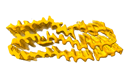
 Downloads & links
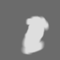
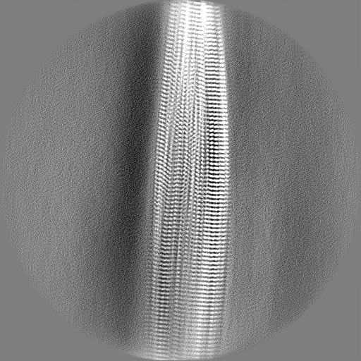
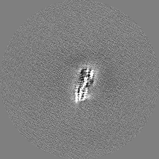
 emd_41611.png
 http://ftp.pdbj.org/pub/emdb/structures/EMD-41611


 Z (Sec.)
 Y (Row.)
 X (Col.)


 Sample components
 Processing
 Electron microscopy
 FIELD EMISSION GUN