- EMDB-41570: Cryo-EM structure of the rat P2X7 receptor in the apo closed state -
+
Open data
ID or keywords:
Loading...
-
Basic information
Entry
Database: EMDB / ID: EMD-41570
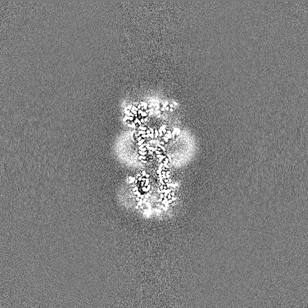
Title
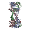
Cryo-EM structure of the rat P2X7 receptor in the apo closed state
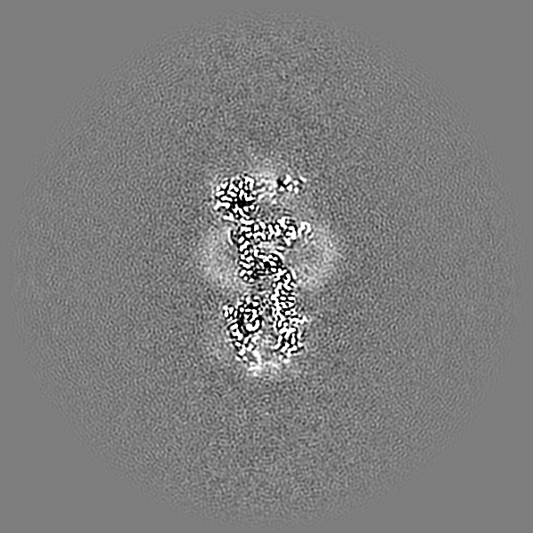
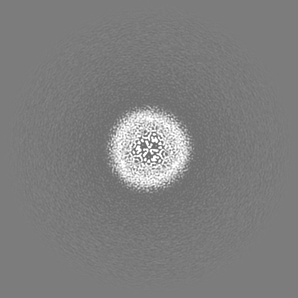
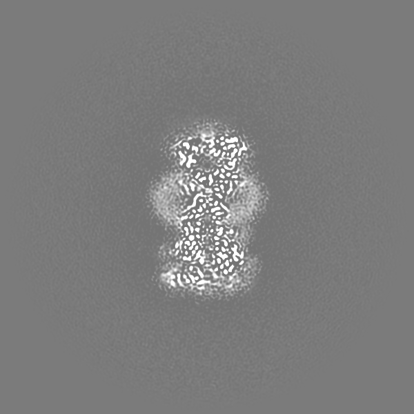
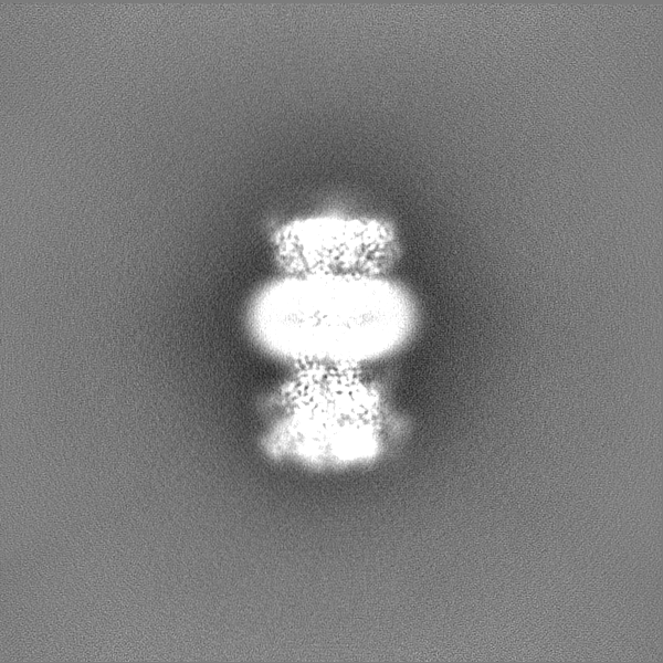
Map data
Sharpened volume of the apo closed state of
Sample
Complex: Membrane protein
Protein or peptide: P2X purinoceptor 7
Ligand: GUANOSINE-5'-DIPHOSPHATE
Ligand: ZINC ION
Ligand: 2-acetamido-2-deoxy-beta-D-glucopyranose
Ligand: PALMITIC ACID
Ligand: SODIUM ION
Ligand: water
Keywords
Membrane Protein / Ion Channel / Ligand-gate Ion Channel / P2X Receptor / Allosteric Antagonist / High-Affinity Agonist
Function / homology
Function and homology information
Platelet homeostasis / The NLRP3 inflammasome / positive regulation of lymphocyte apoptotic process / regulation of presynaptic dense core granule exocytosis / positive regulation of bleb assembly / NAD transport / Elevation of cytosolic Ca2+ levels / phagolysosome assembly / phospholipid transfer to membrane / positive regulation of cytoskeleton organization ...Platelet homeostasis / The NLRP3 inflammasome / positive regulation of lymphocyte apoptotic process / regulation of presynaptic dense core granule exocytosis / positive regulation of bleb assembly / NAD transport / Elevation of cytosolic Ca2+ levels / phagolysosome assembly / phospholipid transfer to membrane / positive regulation of cytoskeleton organization / positive regulation of monoatomic ion transmembrane transport / purinergic nucleotide receptor signaling pathway / plasma membrane organization / extracellularly ATP-gated monoatomic cation channel activity / positive regulation of interleukin-1 alpha production / purinergic nucleotide receptor activity / ATP export / collagen metabolic process / positive regulation of prostaglandin secretion / pore complex assembly / negative regulation of cell volume / plasma membrane phospholipid scrambling / positive regulation of gamma-aminobutyric acid secretion / bleb assembly / vesicle budding from membrane / positive regulation of T cell apoptotic process / bleb / response to fluid shear stress / programmed cell death / positive regulation of ossification / cell volume homeostasis / cellular response to dsRNA / negative regulation of bone resorption / ceramide biosynthetic process / positive regulation of macrophage cytokine production / skeletal system morphogenesis / phospholipid translocation / response to zinc ion / positive regulation of glutamate secretion / sodium channel activity / protein homotrimerization / response to ATP / T cell homeostasis / positive regulation of mitochondrial depolarization / membrane protein ectodomain proteolysis / positive regulation of NLRP3 inflammasome complex assembly / response to electrical stimulus / positive regulation of calcium ion transport into cytosol / synaptic vesicle exocytosis / T cell proliferation / positive regulation of bone mineralization / monoatomic cation transport / potassium channel activity / membrane depolarization / response to mechanical stimulus / regulation of sodium ion transport / neuronal action potential / extrinsic apoptotic signaling pathway / negative regulation of MAPK cascade / release of sequestered calcium ion into cytosol / reactive oxygen species metabolic process / homeostasis of number of cells within a tissue / sensory perception of pain / establishment of localization in cell / positive regulation of glycolytic process / positive regulation of interleukin-1 beta production / protein serine/threonine kinase activator activity / protein catabolic process / positive regulation of protein secretion / response to bacterium / neuromuscular junction / mitochondrion organization / apoptotic signaling pathway / lipopolysaccharide binding / protein processing / response to calcium ion / positive regulation of T cell mediated cytotoxicity / positive regulation of interleukin-6 production / calcium ion transmembrane transport / cell morphogenesis / cell-cell junction / terminal bouton / calcium ion transport / nuclear envelope / channel activity / signaling receptor activity / scaffold protein binding / response to lipopolysaccharide / gene expression / positive regulation of MAPK cascade / cell surface receptor signaling pathway / postsynapse / defense response to Gram-positive bacterium / positive regulation of apoptotic process / response to xenobiotic stimulus / inflammatory response / copper ion binding / signaling receptor binding / external side of plasma membrane / neuronal cell body Similarity search - Function
National Institutes of Health/National Heart, Lung, and Blood Institute (NIH/NHLBI)
R00HL138129
United States
National Institutes of Health/National Institute of General Medical Sciences (NIH/NIGMS)
DP2GM149551
United States
Citation
Journal: Nat Commun / Year: 2024 Title: High-affinity agonism at the P2X receptor is mediated by three residues outside the orthosteric pocket. Authors: Adam C Oken / Nicolas E Lisi / Ipsita Krishnamurthy / Alanna E McCarthy / Michael H Godsey / Arthur Glasfeld / Steven E Mansoor / Abstract: P2X receptors are trimeric ATP-gated ion channels that activate diverse signaling cascades. Due to its role in apoptotic pathways, selective activation of P2X is a potential experimental tool and ...P2X receptors are trimeric ATP-gated ion channels that activate diverse signaling cascades. Due to its role in apoptotic pathways, selective activation of P2X is a potential experimental tool and therapeutic approach in cancer biology. However, mechanisms of high-affinity P2X activation have not been defined. We report high-resolution cryo-EM structures of wild-type rat P2X bound to the high-affinity agonist BzATP as well as significantly improved apo receptor structures in the presence and absence of sodium. Apo structures define molecular details of pore architecture and reveal how a partially hydrated Na ion interacts with the conductance pathway in the closed state. Structural, electrophysiological, and direct binding data of BzATP reveal that three residues just outside the orthosteric ATP-binding site are responsible for its high-affinity agonism. This work provides insights into high-affinity agonism for any P2X receptor and lays the groundwork for development of subtype-specific agonists applicable to cancer therapeutics.
In the structure databanks used in Yorodumi, some data are registered as the other names, "COVID-19 virus" and "2019-nCoV". Here are the details of the virus and the list of structure data.
Jan 31, 2019. EMDB accession codes are about to change! (news from PDBe EMDB page)
EMDB accession codes are about to change! (news from PDBe EMDB page)
The allocation of 4 digits for EMDB accession codes will soon come to an end. Whilst these codes will remain in use, new EMDB accession codes will include an additional digit and will expand incrementally as the available range of codes is exhausted. The current 4-digit format prefixed with “EMD-” (i.e. EMD-XXXX) will advance to a 5-digit format (i.e. EMD-XXXXX), and so on. It is currently estimated that the 4-digit codes will be depleted around Spring 2019, at which point the 5-digit format will come into force.
The EM Navigator/Yorodumi systems omit the EMD- prefix.
Related info.:Q: What is EMD? / ID/Accession-code notation in Yorodumi/EM Navigator
Yorodumi is a browser for structure data from EMDB, PDB, SASBDB, etc.
This page is also the successor to EM Navigator detail page, and also detail information page/front-end page for Omokage search.
The word "yorodu" (or yorozu) is an old Japanese word meaning "ten thousand". "mi" (miru) is to see.
Related info.:EMDB / PDB / SASBDB / Comparison of 3 databanks / Yorodumi Search / Aug 31, 2016. New EM Navigator & Yorodumi / Yorodumi Papers / Jmol/JSmol / Function and homology information / Changes in new EM Navigator and Yorodumi
 Movie
Movie Controller

 Yorodumi
 Open data
 Basic information
 Map data
 Sample
 Keywords
 Function and homology information
 Authors
 United States, 2 items
  Citation
 Structure visualization
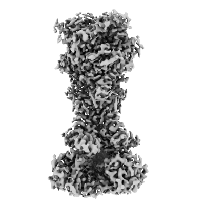
 Downloads & links
 emd_41570.png
 http://ftp.pdbj.org/pub/emdb/structures/EMD-41570
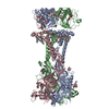


 Z (Sec.)
 Y (Row.)
 X (Col.)


 Sample components
 Homo sapiens (human)


 Processing
 Electron microscopy
 FIELD EMISSION GUN


