ジャーナル: Nat Commun / 年: 2017 タイトル: Dissecting the molecular organization of the translocon-associated protein complex. 著者: Stefan Pfeffer / Johanna Dudek / Miroslava Schaffer / Bobby G Ng / Sahradha Albert / Jürgen M Plitzko / Wolfgang Baumeister / Richard Zimmermann / Hudson H Freeze / Benjamin D Engel / Friedrich Förster / 要旨: In eukaryotic cells, one-third of all proteins must be transported across or inserted into the endoplasmic reticulum (ER) membrane by the ER protein translocon. The translocon-associated protein ...In eukaryotic cells, one-third of all proteins must be transported across or inserted into the endoplasmic reticulum (ER) membrane by the ER protein translocon. The translocon-associated protein (TRAP) complex is an integral component of the translocon, assisting the Sec61 protein-conducting channel by regulating signal sequence and transmembrane helix insertion in a substrate-dependent manner. Here we use cryo-electron tomography (CET) to study the structure of the native translocon in evolutionarily divergent organisms and disease-linked TRAP mutant fibroblasts from human patients. The structural differences detected by subtomogram analysis form a basis for dissecting the molecular organization of the TRAP complex. We assign positions to the four TRAP subunits within the complex, providing insights into their individual functions. The revealed molecular architecture of a central translocon component advances our understanding of membrane protein biogenesis and sheds light on the role of TRAP in human congenital disorders of glycosylation.
 ムービー
ムービー コントローラー


 データを開く
 基本情報
基本情報 マップデータ
 試料
試料
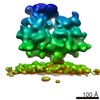
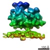
 Chlamydomonas reinhardtii (クラミドモナス)
 データ登録者
 引用
引用


 構造の表示
構造の表示 ムービービューア
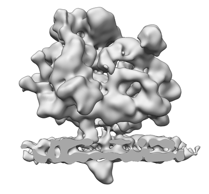
 ダウンロードとリンク
 emd_4145.png
 http://ftp.pdbj.org/pub/emdb/structures/EMD-4145


 Z (Sec.)
 Y (Row.)
 X (Col.)


 試料の構成要素
 解析
解析 電子顕微鏡法
 FIELD EMISSION GUN
