 Movie
Movie Controller


+ Open data

- Basic information
| Entry |  | |||||||||
|---|---|---|---|---|---|---|---|---|---|---|
| Title | Cryotomogram of DIV 4 cultured hippocampal neuron | |||||||||
 Map data Map data | DIV 4 Hippocampal Neuron (Weighted Backprojection) | |||||||||
 Sample Sample |
| |||||||||
 Keywords Keywords | Neuron / Tomogram / denoise / segmentation / CELL ADHESION | |||||||||
| Biological species |  | |||||||||
| Method | electron tomography / cryo EM | |||||||||
 Authors Authors | Swulius MT | |||||||||
| Funding support |  United States, 1 items United States, 1 items
| |||||||||
 Citation Citation | Journal: To be Published Title: Rapid Synthesis of Cryo-ET Data for Training Deep Learning Models. Authors: Purnell C / Heebner J / Hylton RH / Kabonick S / Grillo M / Grigoryev S / Heberle F / Waxham NM / Swulius MT | |||||||||
| History |
|
- Structure visualization
| Supplemental images |
|---|
- Downloads & links
-EMDB archive
| Map data | emd_41256.map.gz | 39 MB |  EMDB map data format EMDB map data format | |
|---|---|---|---|---|
| Header (meta data) | emd-41256-v30.xmlemd-41256.xml | 11.8 KB 11.8 KB | Display Display | EMDB header |
| Images |  emd_41256.png emd_41256.png | 193.8 KB | ||
| Filedesc metadata | emd-41256.cif.gz | 3.4 KB | ||
| Others | emd_41256_additional_1.map.gzemd_41256_additional_2.map.gz | 53.8 MB 688.8 KB | ||
| Archive directory |  http://ftp.pdbj.org/pub/emdb/structures/EMD-41256ftp://ftp.pdbj.org/pub/emdb/structures/EMD-41256 http://ftp.pdbj.org/pub/emdb/structures/EMD-41256ftp://ftp.pdbj.org/pub/emdb/structures/EMD-41256 | HTTPS FTP |
-Related structure data


-Links
| EMDB pages | EMDB (EBI/PDBe) / EMDataResource |
|---|
-Map
| File | Download / File: emd_41256.map.gz / Format: CCP4 / Size: 107.3 MB / Type: IMAGE STORED AS SIGNED BYTE | ||||||||||||||||||||||||||||||||
|---|---|---|---|---|---|---|---|---|---|---|---|---|---|---|---|---|---|---|---|---|---|---|---|---|---|---|---|---|---|---|---|---|---|
| Annotation | DIV 4 Hippocampal Neuron (Weighted Backprojection) | ||||||||||||||||||||||||||||||||
| Projections & slices | Image control
Images are generated by Spider. generated in cubic-lattice coordinate | ||||||||||||||||||||||||||||||||
| Voxel size | X=Y=Z: 13.6 Å | ||||||||||||||||||||||||||||||||
| Density |
| ||||||||||||||||||||||||||||||||
| Symmetry | Space group: 1 | ||||||||||||||||||||||||||||||||
| Details | EMDB XML:
|
 Z (Sec.)
 Y (Row.)
 X (Col.)


-Supplemental data
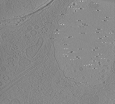
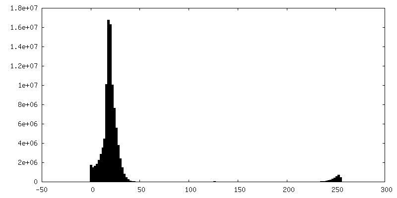
-Additional map: DIV 4 Hippocampal Neuron (regression denoised)
| File | emd_41256_additional_1.map | ||||||||||||
|---|---|---|---|---|---|---|---|---|---|---|---|---|---|
| Annotation | DIV 4 Hippocampal Neuron (regression denoised) | ||||||||||||
| Projections & Slices |
| ||||||||||||
| Density Histograms |


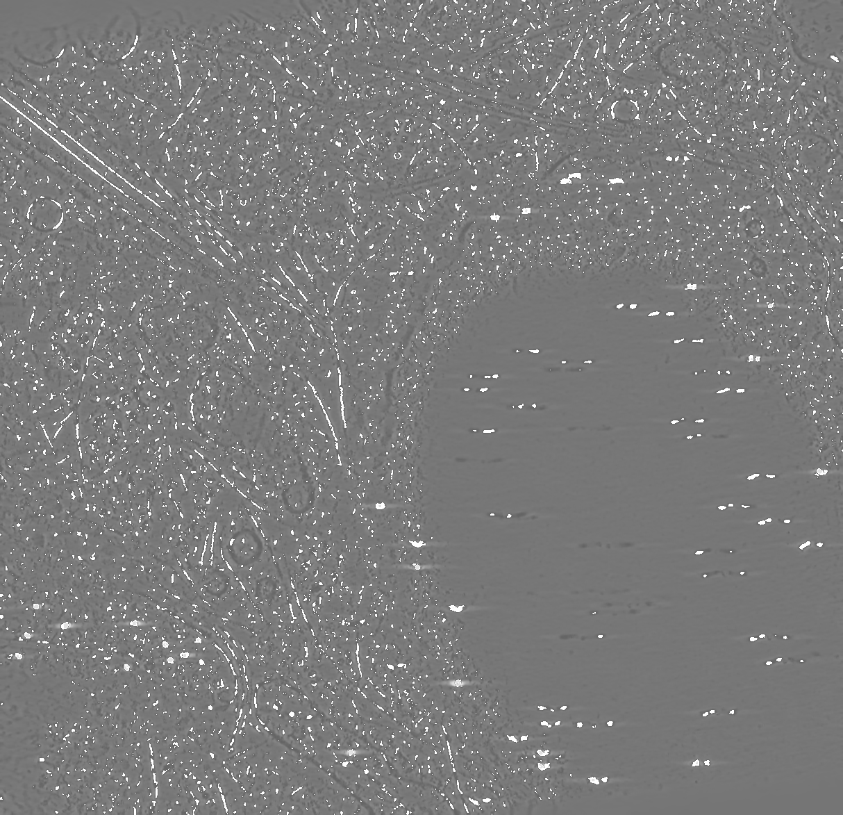


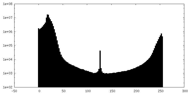
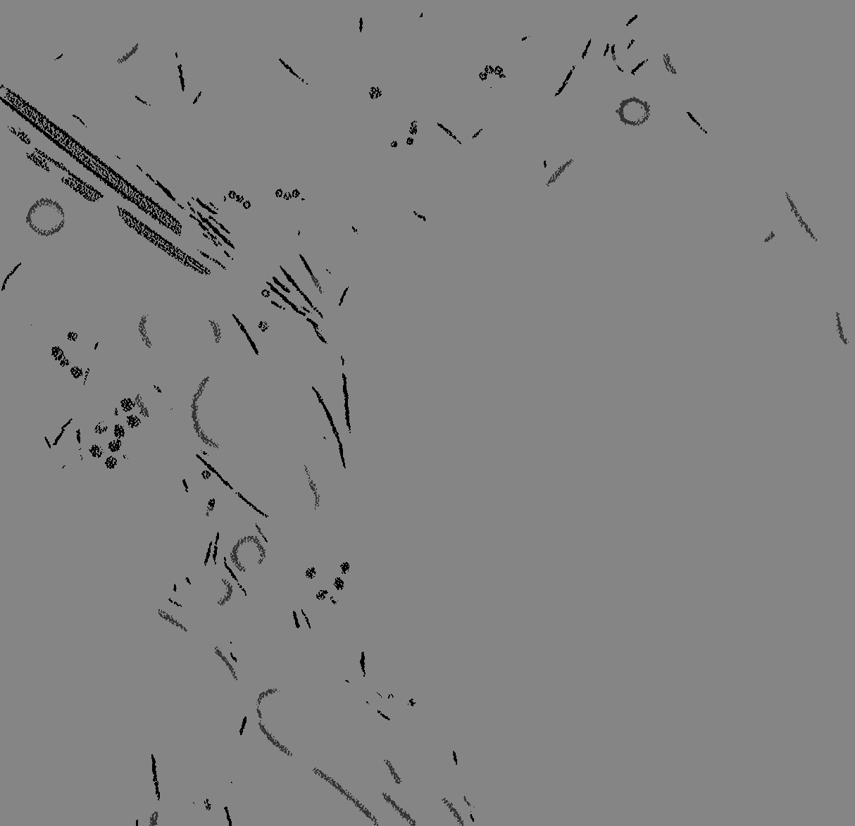
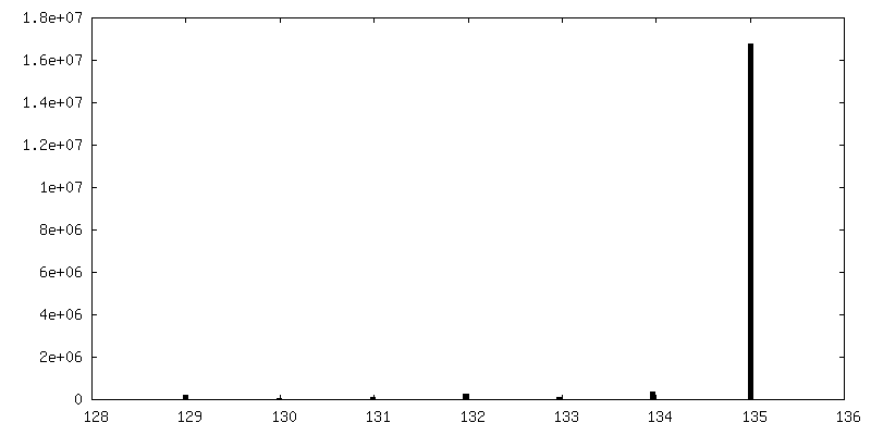
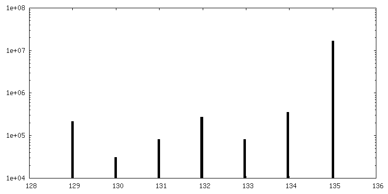
-Additional map: DIV 4 Hippocampal Neuron (Greyscale Segmentation)
| File | emd_41256_additional_2.map | ||||||||||||
|---|---|---|---|---|---|---|---|---|---|---|---|---|---|
| Annotation | DIV 4 Hippocampal Neuron (Greyscale Segmentation) | ||||||||||||
| Projections & Slices |
| ||||||||||||
| Density Histograms |


- Sample components
-Entire : DIV 4 Hippocampal Neuron
| Entire | Name: DIV 4 Hippocampal Neuron |
|---|---|
| Components |
|
-Supramolecule #1: DIV 4 Hippocampal Neuron
| Supramolecule | Name: DIV 4 Hippocampal Neuron / type: cell / ID: 1 / Parent: 0 Details: Weighted Backprojection, Regression Denoised, and Greyscale Segmentation |
|---|---|
| Source (natural) | Organism: |
-Experimental details
-Structure determination
| Method | cryo EM |
|---|---|
 Processing Processing | electron tomography |
| Aggregation state | cell |
-Sample preparation
| Buffer | pH: 7 |
|---|---|
| Vitrification | Cryogen name: ETHANE |
| Sectioning | Other: NO SECTIONING |
- Electron microscopy
| Microscope | TFS KRIOS |
|---|---|
| Image recording | Film or detector model: GATAN K3 BIOQUANTUM (6k x 4k) / Average electron dose: 2.0 e/Å2 |
| Electron beam | Acceleration voltage: 300 kV / Electron source:  FIELD EMISSION GUN FIELD EMISSION GUN |
| Electron optics | Illumination mode: FLOOD BEAM / Imaging mode: BRIGHT FIELD / Cs: 2.7 mm / Nominal defocus max: 8.0 µm / Nominal defocus min: 8.0 µm |
| Experimental equipment |  Model: Titan Krios / Image courtesy: FEI Company |
-Image processing
| Final reconstruction | Number images used: 60 |
|---|
