Journal: Elife / Year: 2016 Title: Accelerated cryo-EM structure determination with parallelisation using GPUs in RELION-2. Authors: Dari Kimanius / Björn O Forsberg / Sjors Hw Scheres / Erik Lindahl / Abstract: By reaching near-atomic resolution for a wide range of specimens, single-particle cryo-EM structure determination is transforming structural biology. However, the necessary calculations come at large ...By reaching near-atomic resolution for a wide range of specimens, single-particle cryo-EM structure determination is transforming structural biology. However, the necessary calculations come at large computational costs, which has introduced a bottleneck that is currently limiting throughput and the development of new methods. Here, we present an implementation of the RELION image processing software that uses graphics processors (GPUs) to address the most computationally intensive steps of its cryo-EM structure determination workflow. Both image classification and high-resolution refinement have been accelerated more than an order-of-magnitude, and template-based particle selection has been accelerated well over two orders-of-magnitude on desktop hardware. Memory requirements on GPUs have been reduced to fit widely available hardware, and we show that the use of single precision arithmetic does not adversely affect results. This enables high-resolution cryo-EM structure determination in a matter of days on a single workstation.
History
Deposition
Sep 21, 2016
-
Header (metadata) release
Oct 5, 2016
-
Map release
Oct 5, 2016
-
Update
Jul 29, 2020
-
Current status
Jul 29, 2020
Processing site: PDBe / Status: Released
-
Structure visualization
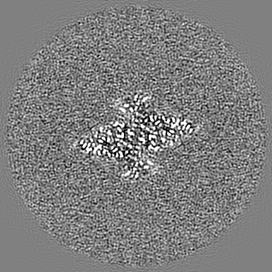
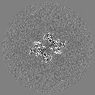
Movie
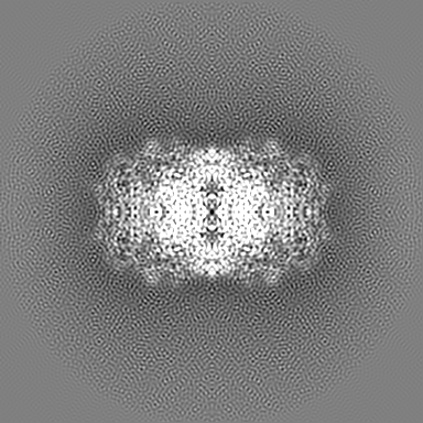
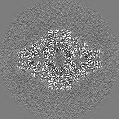
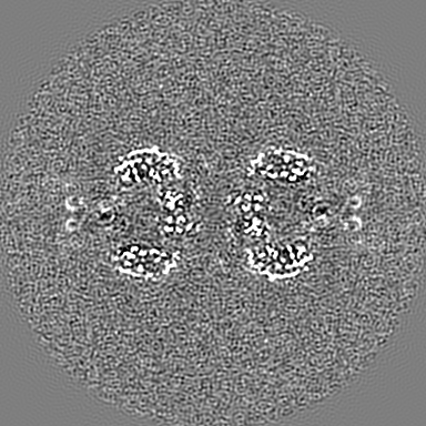
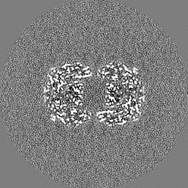
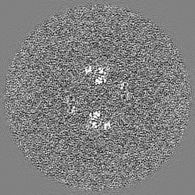
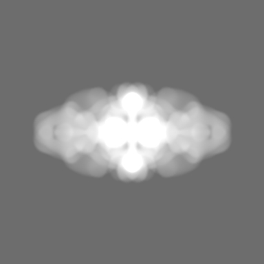
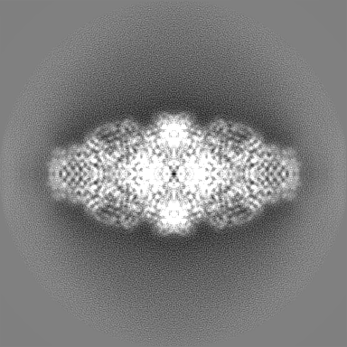
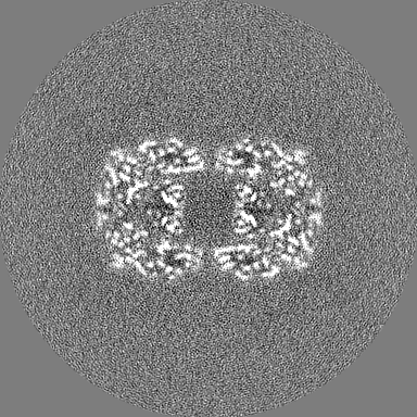
Surface view with section colored by density value
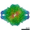
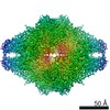
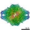
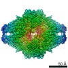
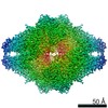
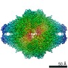
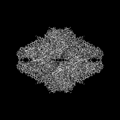
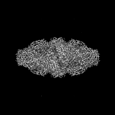
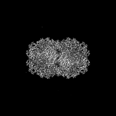
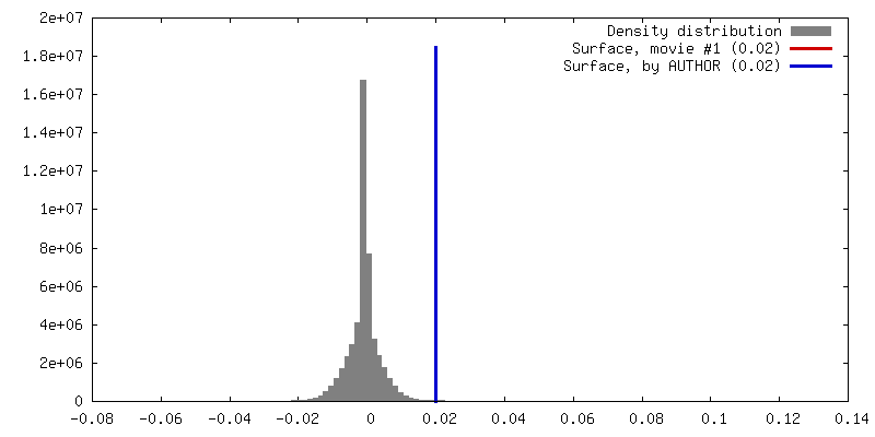
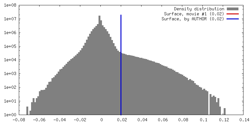
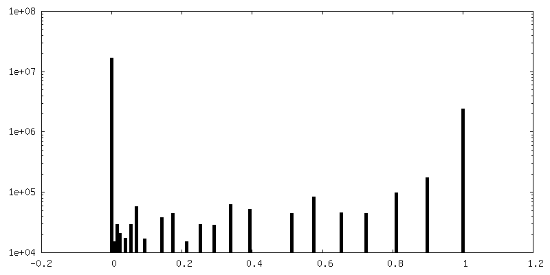
Number classes used: 1 / Applied symmetry - Point group: D2 (2x2 fold dihedral) / Algorithm: FOURIER SPACE / Resolution.type: BY AUTHOR / Resolution: 2.2 Å / Resolution method: FSC 0.143 CUT-OFF / Software - Name: RELION (ver. 2.0) Details: Standard RELION post-processing, i.e. convolution effects of the solvent mask are corrected for using phase-randomisation. Number images used: 108209
Initial angle assignment
Type: PROJECTION MATCHING / Software - Name: RELION (ver. 2.0) / Details: Standard RELION 3D auto-refine
Final angle assignment
Type: PROJECTION MATCHING / Software - Name: RELION (ver. 2.0) / Details: Standard RELION 3D auto-refine
Final 3D classification
Number classes: 8 / Software - Name: RELION (ver. 2.0) Details: Selected 7 out of 8 3D classes. Also 2D classification was performed to select suitable particles.
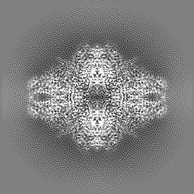
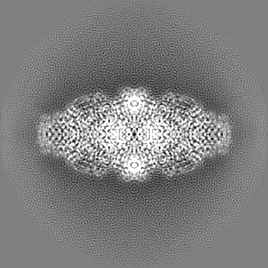
FSC plot (resolution estimation)
+
About Yorodumi
-
News
-
Feb 9, 2022. New format data for meta-information of EMDB entries
New format data for meta-information of EMDB entries
Version 3 of the EMDB header file is now the official format.
The previous official version 1.9 will be removed from the archive.
In the structure databanks used in Yorodumi, some data are registered as the other names, "COVID-19 virus" and "2019-nCoV". Here are the details of the virus and the list of structure data.
Jan 31, 2019. EMDB accession codes are about to change! (news from PDBe EMDB page)
EMDB accession codes are about to change! (news from PDBe EMDB page)
The allocation of 4 digits for EMDB accession codes will soon come to an end. Whilst these codes will remain in use, new EMDB accession codes will include an additional digit and will expand incrementally as the available range of codes is exhausted. The current 4-digit format prefixed with “EMD-” (i.e. EMD-XXXX) will advance to a 5-digit format (i.e. EMD-XXXXX), and so on. It is currently estimated that the 4-digit codes will be depleted around Spring 2019, at which point the 5-digit format will come into force.
The EM Navigator/Yorodumi systems omit the EMD- prefix.
Related info.:Q: What is EMD? / ID/Accession-code notation in Yorodumi/EM Navigator
Yorodumi is a browser for structure data from EMDB, PDB, SASBDB, etc.
This page is also the successor to EM Navigator detail page, and also detail information page/front-end page for Omokage search.
The word "yorodu" (or yorozu) is an old Japanese word meaning "ten thousand". "mi" (miru) is to see.
Related info.:EMDB / PDB / SASBDB / Comparison of 3 databanks / Yorodumi Search / Aug 31, 2016. New EM Navigator & Yorodumi / Yorodumi Papers / Jmol/JSmol / Function and homology information / Changes in new EM Navigator and Yorodumi
 Movie
Movie Controller

 Yorodumi
 Open data
 Basic information
 Map data
 Sample
 Function and homology information
 Authors
 Citation

 Structure visualization
 Downloads & links
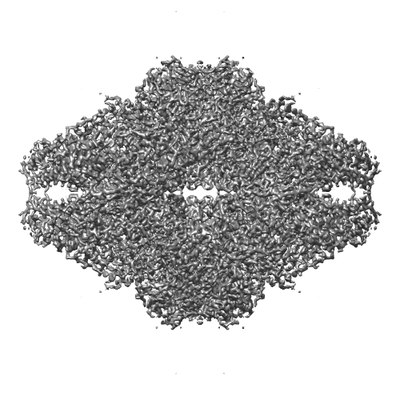 emd_4116.png
 http://ftp.pdbj.org/pub/emdb/structures/EMD-4116


 Z (Sec.)
 Y (Row.)
 X (Col.)


 Sample components
 Processing
 Electron microscopy
 FIELD EMISSION GUN

