+ Open data
- Basic information
| Entry |  | |||||||||||||||
|---|---|---|---|---|---|---|---|---|---|---|---|---|---|---|---|---|
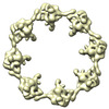
| Title | Cytoplasmic outer ring of the yeast NPC | |||||||||||||||
 Map data Map data | c8 map of cytoplasmic outer ring reconstructed from multibody volume | |||||||||||||||
 Sample Sample |
| |||||||||||||||
 Keywords Keywords | nuclear pore complex / nucleocytoplasmic transport / nucleoporin / membrane protein / TRANSPORT PROTEIN | |||||||||||||||
| Biological species |  | |||||||||||||||
| Method | single particle reconstruction / cryo EM / Resolution: 22.0 Å | |||||||||||||||
 Authors Authors | Akey CW / Echeverria I / Ouch C / Fernandez-Martinez J / Rout MP | |||||||||||||||
| Funding support |  United States, 4 items United States, 4 items
| |||||||||||||||
 Citation Citation |  Journal: Mol Cell / Year: 2023 Journal: Mol Cell / Year: 2023Title: Implications of a multiscale structure of the yeast nuclear pore complex. Authors: Christopher W Akey / Ignacia Echeverria / Christna Ouch / Ilona Nudelman / Yi Shi / Junjie Wang / Brian T Chait / Andrej Sali / Javier Fernandez-Martinez / Michael P Rout /   Abstract: Nuclear pore complexes (NPCs) direct the nucleocytoplasmic transport of macromolecules. Here, we provide a composite multiscale structure of the yeast NPC, based on improved 3D density maps from ...Nuclear pore complexes (NPCs) direct the nucleocytoplasmic transport of macromolecules. Here, we provide a composite multiscale structure of the yeast NPC, based on improved 3D density maps from cryogenic electron microscopy and AlphaFold2 models. Key features of the inner and outer rings were integrated into a comprehensive model. We resolved flexible connectors that tie together the core scaffold, along with equatorial transmembrane complexes and a lumenal ring that anchor this channel within the pore membrane. The organization of the nuclear double outer ring reveals an architecture that may be shared with ancestral NPCs. Additional connections between the core scaffold and the central transporter suggest that under certain conditions, a degree of local organization is present at the periphery of the transport machinery. These connectors may couple conformational changes in the scaffold to the central transporter to modulate transport. Collectively, this analysis provides insights into assembly, transport, and NPC evolution. | |||||||||||||||
| History |
|
- Structure visualization
| Supplemental images |
|---|
- Downloads & links
-EMDB archive
| Map data |  emd_41119.map.gz emd_41119.map.gz | 23.8 MB |  EMDB map data format EMDB map data format | |
|---|---|---|---|---|
| Header (meta data) |  emd-41119-v30.xml emd-41119-v30.xml emd-41119.xml emd-41119.xml | 27.6 KB 27.6 KB | Display Display |  EMDB header EMDB header |
| FSC (resolution estimation) |  emd_41119_fsc.xml emd_41119_fsc.xml | 21.9 KB | Display |  FSC data file FSC data file |
| Images |  emd_41119.png emd_41119.png | 126.7 KB | ||
| Filedesc metadata |  emd-41119.cif.gz emd-41119.cif.gz | 5.9 KB | ||
| Others |  emd_41119_additional_1.map.gz emd_41119_additional_1.map.gz emd_41119_additional_2.map.gz emd_41119_additional_2.map.gz emd_41119_additional_3.map.gz emd_41119_additional_3.map.gz emd_41119_half_map_1.map.gz emd_41119_half_map_1.map.gz emd_41119_half_map_2.map.gz emd_41119_half_map_2.map.gz | 10 MB 9.9 MB 38.2 MB 23.3 MB 23.3 MB | ||
| Archive directory |  http://ftp.pdbj.org/pub/emdb/structures/EMD-41119 http://ftp.pdbj.org/pub/emdb/structures/EMD-41119 ftp://ftp.pdbj.org/pub/emdb/structures/EMD-41119 ftp://ftp.pdbj.org/pub/emdb/structures/EMD-41119 | HTTPS FTP |
-Validation report
| Summary document |  emd_41119_validation.pdf.gz emd_41119_validation.pdf.gz | 493.3 KB | Display |  EMDB validaton report EMDB validaton report |
|---|---|---|---|---|
| Full document |  emd_41119_full_validation.pdf.gz emd_41119_full_validation.pdf.gz | 492.8 KB | Display | |
| Data in XML |  emd_41119_validation.xml.gz emd_41119_validation.xml.gz | 24.5 KB | Display | |
| Data in CIF |  emd_41119_validation.cif.gz emd_41119_validation.cif.gz | 33.1 KB | Display | |
| Arichive directory |  https://ftp.pdbj.org/pub/emdb/validation_reports/EMD-41119 https://ftp.pdbj.org/pub/emdb/validation_reports/EMD-41119 ftp://ftp.pdbj.org/pub/emdb/validation_reports/EMD-41119 ftp://ftp.pdbj.org/pub/emdb/validation_reports/EMD-41119 | HTTPS FTP |
-Related structure data
- Links
Links
| EMDB pages |  EMDB (EBI/PDBe) / EMDB (EBI/PDBe) /  EMDataResource EMDataResource |
|---|---|
| Related items in Molecule of the Month |
- Map
Map
| File |  Download / File: emd_41119.map.gz / Format: CCP4 / Size: 421.9 MB / Type: IMAGE STORED AS FLOATING POINT NUMBER (4 BYTES) Download / File: emd_41119.map.gz / Format: CCP4 / Size: 421.9 MB / Type: IMAGE STORED AS FLOATING POINT NUMBER (4 BYTES) | ||||||||||||||||||||||||||||||||||||
|---|---|---|---|---|---|---|---|---|---|---|---|---|---|---|---|---|---|---|---|---|---|---|---|---|---|---|---|---|---|---|---|---|---|---|---|---|---|
| Annotation | c8 map of cytoplasmic outer ring reconstructed from multibody volume | ||||||||||||||||||||||||||||||||||||
| Projections & slices | Image control
Images are generated by Spider. | ||||||||||||||||||||||||||||||||||||
| Voxel size | X=Y=Z: 2.66 Å | ||||||||||||||||||||||||||||||||||||
| Density |
| ||||||||||||||||||||||||||||||||||||
| Symmetry | Space group: 1 | ||||||||||||||||||||||||||||||||||||
| Details | EMDB XML:
|
-Supplemental data
-Additional map: half map of cytoplasmic outer ring multibody volume
| File | emd_41119_additional_1.map | ||||||||||||
|---|---|---|---|---|---|---|---|---|---|---|---|---|---|
| Annotation | half map of cytoplasmic outer ring multibody volume | ||||||||||||
| Projections & Slices |
| ||||||||||||
| Density Histograms |
-Additional map: half map of cytoplasmic outer ring multibody volume
| File | emd_41119_additional_2.map | ||||||||||||
|---|---|---|---|---|---|---|---|---|---|---|---|---|---|
| Annotation | half map of cytoplasmic outer ring multibody volume | ||||||||||||
| Projections & Slices |
| ||||||||||||
| Density Histograms |
-Additional map: 2.5x multibody map of cytoplasmic outer ring
| File | emd_41119_additional_3.map | ||||||||||||
|---|---|---|---|---|---|---|---|---|---|---|---|---|---|
| Annotation | 2.5x multibody map of cytoplasmic outer ring | ||||||||||||
| Projections & Slices |
| ||||||||||||
| Density Histograms |
-Half map: half map of c8 cytoplasmic outer ring
| File | emd_41119_half_map_1.map | ||||||||||||
|---|---|---|---|---|---|---|---|---|---|---|---|---|---|
| Annotation | half map of c8 cytoplasmic outer ring | ||||||||||||
| Projections & Slices |
| ||||||||||||
| Density Histograms |
-Half map: half map of c8 cytoplasmic outer ring
| File | emd_41119_half_map_2.map | ||||||||||||
|---|---|---|---|---|---|---|---|---|---|---|---|---|---|
| Annotation | half map of c8 cytoplasmic outer ring | ||||||||||||
| Projections & Slices |
| ||||||||||||
| Density Histograms |
- Sample components
-Entire : Nuclear Pore Complex
| Entire | Name: Nuclear Pore Complex |
|---|---|
| Components |
|
-Supramolecule #1: Nuclear Pore Complex
| Supramolecule | Name: Nuclear Pore Complex / type: complex / ID: 1 / Parent: 0 / Details: Protein A tagged Mlp1 pullout of NPC |
|---|---|
| Source (natural) | Organism:  Strain: MATa ade2-1 ura3-1 his3-11,15 trp1-1 leu2-3,112 can1-100 MLP1-PPX-ProteinA::HIS5 Organelle: nucleus / Location in cell: nuclear envelope |
-Experimental details
-Structure determination
| Method | cryo EM |
|---|---|
 Processing Processing | single particle reconstruction |
| Aggregation state | particle |
- Sample preparation
| Concentration | 0.1 mg/mL |
|---|---|
| Buffer | pH: 7.5 Details: 20mM HEPES,50mM Potassium acetate,20mM NaCl,2mM MgCl2,1mM DTT |
| Grid | Model: Quantifoil R2/2 / Material: COPPER / Mesh: 400 / Support film - Material: CARBON / Support film - topology: HOLEY ARRAY / Support film - Film thickness: 5 / Pretreatment - Type: GLOW DISCHARGE / Pretreatment - Time: 30 sec. / Pretreatment - Atmosphere: OTHER |
| Vitrification | Cryogen name: ETHANE / Chamber humidity: 100 % / Chamber temperature: 283 K / Instrument: FEI VITROBOT MARK III |
| Details | One step affinity purified |
- Electron microscopy
| Microscope | FEI TITAN KRIOS |
|---|---|
| Specialist optics | Energy filter - Name: GIF Bioquantum / Energy filter - Slit width: 20 eV |
| Details | Preliminary grid screening done manually with individual images of low magnification montages of candidate meshes. |
| Image recording | Film or detector model: GATAN K2 SUMMIT (4k x 4k) / Detector mode: SUPER-RESOLUTION / Digitization - Dimensions - Width: 3838 pixel / Digitization - Dimensions - Height: 3710 pixel / Digitization - Frames/image: 2-40 / Number grids imaged: 1 / Number real images: 4015 / Average electron dose: 40.0 e/Å2 / Details: 3218 images retained after triage |
| Electron beam | Acceleration voltage: 300 kV / Electron source:  FIELD EMISSION GUN FIELD EMISSION GUN |
| Electron optics | C2 aperture diameter: 100.0 µm / Calibrated magnification: 37651 / Illumination mode: FLOOD BEAM / Imaging mode: BRIGHT FIELD / Cs: 2.7 mm / Nominal defocus max: 3.5 µm / Nominal defocus min: 1.5 µm |
| Sample stage | Specimen holder model: FEI TITAN KRIOS AUTOGRID HOLDER / Cooling holder cryogen: NITROGEN |
| Experimental equipment |  Model: Titan Krios / Image courtesy: FEI Company |
+ Image processing
-Atomic model buiding 1
| Initial model | Chain - Source name: Other / Chain - Initial model type: experimental model / Details: none |
|---|---|
| Details | Rigid body docking of 3D map of cytoplasmic outer ring into NPC 3D map with Chimera |
| Refinement | Space: REAL / Protocol: RIGID BODY FIT / Target criteria: cross correlation |
 Movie
Movie Controller


 Z (Sec.)
 Y (Row.)
 X (Col.)


