National Institutes of Health/National Heart, Lung, and Blood Institute (NIH/NHLBI)
DP5-OD019800
United States
Citation
Journal: Nat Struct Mol Biol / Year: 2023 Title: Structure of the preholoproteasome reveals late steps in proteasome core particle biogenesis. Authors: Richard M Walsh / Shaun Rawson / Helena M Schnell / Benjamin Velez / Tamayanthi Rajakumar / John Hanna / Abstract: Assembly of the proteasome's core particle (CP), a barrel-shaped chamber of four stacked rings, requires five chaperones and five subunit propeptides. Fusion of two half-CP precursors yields a ...Assembly of the proteasome's core particle (CP), a barrel-shaped chamber of four stacked rings, requires five chaperones and five subunit propeptides. Fusion of two half-CP precursors yields a complete structure but remains immature until active site maturation. Here, using Saccharomyces cerevisiae, we report a high-resolution cryogenic electron microscopy structure of preholoproteasome, a post-fusion assembly intermediate. Our data reveal how CP midline-spanning interactions induce local changes in structure, facilitating maturation. Unexpectedly, we find that cleavage may not be sufficient for propeptide release, as residual interactions with chaperones such as Ump1 hold them in place. We evaluated previous models proposing that dynamic conformational changes in chaperones drive CP fusion and autocatalytic activation by comparing preholoproteasome to pre-fusion intermediates. Instead, the data suggest a scaffolding role for the chaperones Ump1 and Pba1/Pba2. Our data clarify key aspects of CP assembly, suggest that undiscovered mechanisms exist to explain CP fusion/activation, and have relevance for diseases of defective CP biogenesis.
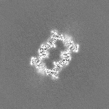
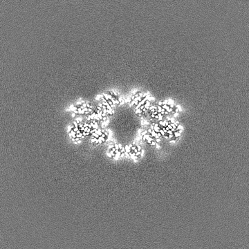
Name: Proteasome 20S core particle from Pre1-1 Pre4-1 Double mutant type: complex / ID: 1 / Parent: 0 / Macromolecule list: all Details: Pre1-1 mutation is S142F in Proteasome subunit beta type-4. Pre4-1 Mutation is the deletion of the last 15 residues from the C-terminus of Proteasome subunit beta type-7.
Cryogen name: ETHANE / Chamber humidity: 100 % / Chamber temperature: 295.15 K / Instrument: FEI VITROBOT MARK IV
-
Electron microscopy
Microscope
FEI TITAN KRIOS
Specialist optics
Energy filter - Name: GIF Bioquantum / Energy filter - Slit width: 25 eV
Image recording
Film or detector model: GATAN K3 BIOQUANTUM (6k x 4k) / Number grids imaged: 2 / Number real images: 33116 / Average exposure time: 3.8 sec. / Average electron dose: 54.3 e/Å2
Electron beam
Acceleration voltage: 300 kV / Electron source: FIELD EMISSION GUN
In the structure databanks used in Yorodumi, some data are registered as the other names, "COVID-19 virus" and "2019-nCoV". Here are the details of the virus and the list of structure data.
Jan 31, 2019. EMDB accession codes are about to change! (news from PDBe EMDB page)
EMDB accession codes are about to change! (news from PDBe EMDB page)
The allocation of 4 digits for EMDB accession codes will soon come to an end. Whilst these codes will remain in use, new EMDB accession codes will include an additional digit and will expand incrementally as the available range of codes is exhausted. The current 4-digit format prefixed with “EMD-” (i.e. EMD-XXXX) will advance to a 5-digit format (i.e. EMD-XXXXX), and so on. It is currently estimated that the 4-digit codes will be depleted around Spring 2019, at which point the 5-digit format will come into force.
The EM Navigator/Yorodumi systems omit the EMD- prefix.
Related info.:Q: What is EMD? / ID/Accession-code notation in Yorodumi/EM Navigator
Yorodumi is a browser for structure data from EMDB, PDB, SASBDB, etc.
This page is also the successor to EM Navigator detail page, and also detail information page/front-end page for Omokage search.
The word "yorodu" (or yorozu) is an old Japanese word meaning "ten thousand". "mi" (miru) is to see.
Related info.:EMDB / PDB / SASBDB / Comparison of 3 databanks / Yorodumi Search / Aug 31, 2016. New EM Navigator & Yorodumi / Yorodumi Papers / Jmol/JSmol / Function and homology information / Changes in new EM Navigator and Yorodumi
 Movie
Movie Controller


 Open data
 Basic information
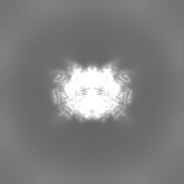
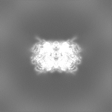
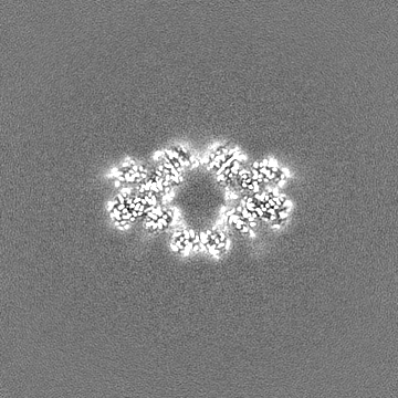
 Map data
 Sample
 Keywords
 Function and homology information
 Authors
 United States, 1 items
  Citation
 Structure visualization
 Downloads & links
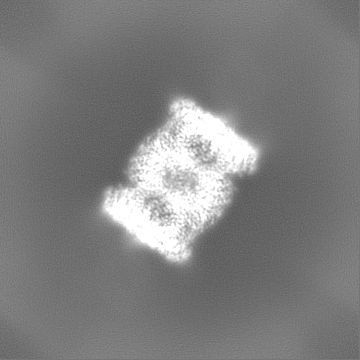
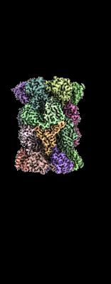 emd_40944.png
 http://ftp.pdbj.org/pub/emdb/structures/EMD-40944


 Z (Sec.)
 Y (Row.)
 X (Col.)


 Sample components
 Processing
 Electron microscopy
 FIELD EMISSION GUN

