 Movie
Movie Controller


+ Open data

- Basic information
| Entry |  | |||||||||
|---|---|---|---|---|---|---|---|---|---|---|
| Title | ExoKCR1 channelrhodopsin | |||||||||
 Map data Map data | ||||||||||
 Sample Sample |
| |||||||||
 Keywords Keywords | Channelrhodopsin / MEMBRANE PROTEIN | |||||||||
| Biological species |  Hyphochytrium catenoides (eukaryote) Hyphochytrium catenoides (eukaryote) | |||||||||
| Method | single particle reconstruction / cryo EM / Resolution: 2.6 Å | |||||||||
 Authors Authors | Zhang MF | |||||||||
| Funding support | 1 items
| |||||||||
 Citation Citation | Journal: Nat Commun / Year: 2024 Title: Channelrhodopsins with distinct chromophores and binding patterns. Authors: Yuanyue Shan / Liping Zhao / Meiyu Chen / Xiao Li / Mingfeng Zhang / Duanqing Pei /  Abstract: Channelrhodopsins are popular optogenetic tools in neuroscience, but remain poorly understood mechanistically. Here we report the cryo-EM structures of channelrhodopsin-2 (ChR2) from Chlamydomonas ...Channelrhodopsins are popular optogenetic tools in neuroscience, but remain poorly understood mechanistically. Here we report the cryo-EM structures of channelrhodopsin-2 (ChR2) from Chlamydomonas reinhardtii and H. catenoides kalium channelrhodopsin (KCR1). We show that ChR2 recruits an endogenous N-retinylidene-PE-like molecule to a previously unidentified lateral retinal binding pocket, exhibiting a reduced light response in HEK293 cells. In contrast, H. catenoides kalium channelrhodopsin (KCR1) binds an endogenous retinal in its canonical retinal binding pocket under identical condition. However, exogenous ATR reduces the photocurrent magnitude of wild type KCR1 and also inhibits its leaky mutant C110T. Our results uncover diverse retinal chromophores with distinct binding patterns for channelrhodopsins in mammalian cells, which may further inspire next generation optogenetics for complex tasks such as cell fate control. | |||||||||
| History |
|
- Structure visualization
| Supplemental images |
|---|

- Downloads & links
-EMDB archive
| Map data | emd_39882.map.gz | 943.8 MB |  EMDB map data format EMDB map data format | |
|---|---|---|---|---|
| Header (meta data) | emd-39882-v30.xmlemd-39882.xml | 13.4 KB 13.4 KB | Display Display | EMDB header |
| Images |  emd_39882.png emd_39882.png | 39.8 KB | ||
| Filedesc metadata | emd-39882.cif.gz | 5.2 KB | ||
| Others | emd_39882_half_map_1.map.gzemd_39882_half_map_2.map.gz | 927.3 MB 927.3 MB | ||
| Archive directory |  http://ftp.pdbj.org/pub/emdb/structures/EMD-39882ftp://ftp.pdbj.org/pub/emdb/structures/EMD-39882 http://ftp.pdbj.org/pub/emdb/structures/EMD-39882ftp://ftp.pdbj.org/pub/emdb/structures/EMD-39882 | HTTPS FTP |
-Related structure data
| Related structure data |  8zaoMC  8zamC 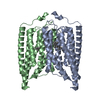 8zanC  8zapC  8zaqC M: atomic model generated by this map C: citing same article ( |
|---|


-Links
| EMDB pages | EMDB (EBI/PDBe) / EMDataResource |
|---|
-Map
| File | Download / File: emd_39882.map.gz / Format: CCP4 / Size: 1000 MB / Type: IMAGE STORED AS FLOATING POINT NUMBER (4 BYTES) | ||||||||||||||||||||||||||||||||||||
|---|---|---|---|---|---|---|---|---|---|---|---|---|---|---|---|---|---|---|---|---|---|---|---|---|---|---|---|---|---|---|---|---|---|---|---|---|---|
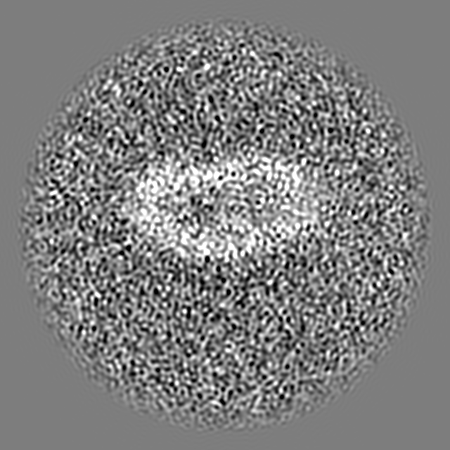
| Projections & slices | Image control
Images are generated by Spider. | ||||||||||||||||||||||||||||||||||||
| Voxel size | X=Y=Z: 0.35 Å | ||||||||||||||||||||||||||||||||||||
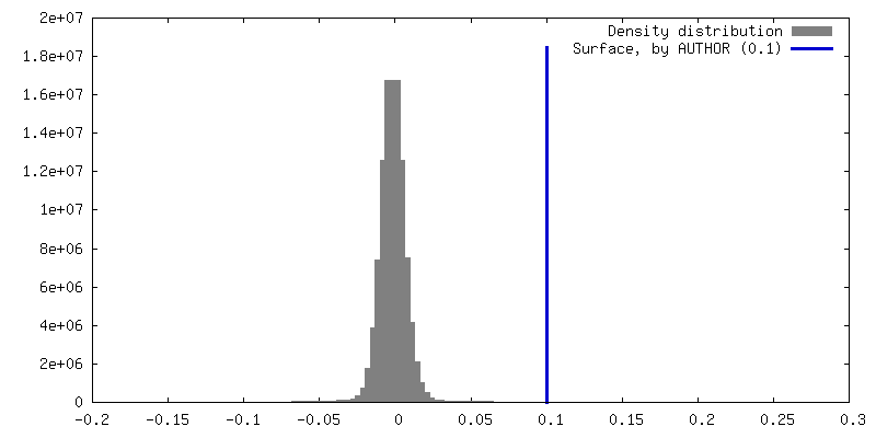
| Density |
| ||||||||||||||||||||||||||||||||||||
| Symmetry | Space group: 1 | ||||||||||||||||||||||||||||||||||||
| Details | EMDB XML:
|
 Z (Sec.)
 Y (Row.)
 X (Col.)


-Supplemental data
-Half map: #2
| File | emd_39882_half_map_1.map | ||||||||||||
|---|---|---|---|---|---|---|---|---|---|---|---|---|---|
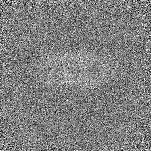
| Projections & Slices |
| ||||||||||||
| Density Histograms |


-Half map: #1
| File | emd_39882_half_map_2.map | ||||||||||||
|---|---|---|---|---|---|---|---|---|---|---|---|---|---|
| Projections & Slices |
| ||||||||||||
| Density Histograms |


- Sample components
-Entire : exoKCR1
| Entire | Name: exoKCR1 |
|---|---|
| Components |
|
-Supramolecule #1: exoKCR1
| Supramolecule | Name: exoKCR1 / type: complex / ID: 1 / Parent: 0 / Macromolecule list: #1 |
|---|---|
| Source (natural) | Organism: Hyphochytrium catenoides (eukaryote) |
-Macromolecule #1: exoKCR1
| Macromolecule | Name: exoKCR1 / type: protein_or_peptide / ID: 1 / Number of copies: 3 / Enantiomer: LEVO |
|---|---|
| Source (natural) | Organism: Hyphochytrium catenoides (eukaryote) |
| Molecular weight | Theoretical: 29.856984 KDa |
| Recombinant expression | Organism:  Homo sapiens (human) Homo sapiens (human) |
| Sequence | String: PFYDSRPPEG WPKGSINDMD YPLLGSICAV CCVFVAGSGI WMLYRLDLGM GYSCKPYKSG RAPEVNSLSG IICLLCGTMY AAKSFDFFD GGGTPFSLNW YWYLDYVFTC PLLILDFAFT LDLPHKIRYF FAVFLTLWCG VAAFVTPSAY RFAYYALGCC W FTPFALSL ...String: PFYDSRPPEG WPKGSINDMD YPLLGSICAV CCVFVAGSGI WMLYRLDLGM GYSCKPYKSG RAPEVNSLSG IICLLCGTMY AAKSFDFFD GGGTPFSLNW YWYLDYVFTC PLLILDFAFT LDLPHKIRYF FAVFLTLWCG VAAFVTPSAY RFAYYALGCC W FTPFALSL MRHVKERYLV YPPKCQRWLF WACVIFFGFW PMFPILFIFS WLGTGHISQQ AFYIIHAFLD LTCKSIFGIL MT VFRLELE EHTEVQGLPL NE |
-Macromolecule #2: POTASSIUM ION
| Macromolecule | Name: POTASSIUM ION / type: ligand / ID: 2 / Number of copies: 15 / Formula: K |
|---|---|
| Molecular weight | Theoretical: 39.098 Da |
-Macromolecule #3: RETINAL
| Macromolecule | Name: RETINAL / type: ligand / ID: 3 / Number of copies: 3 / Formula: RET |
|---|---|
| Molecular weight | Theoretical: 284.436 Da |
| Chemical component information |  ChemComp-RET: |
-Macromolecule #4: water
| Macromolecule | Name: water / type: ligand / ID: 4 / Number of copies: 30 / Formula: HOH |
|---|---|
| Molecular weight | Theoretical: 18.015 Da |
| Chemical component information |  ChemComp-HOH: |
-Experimental details
-Structure determination
| Method | cryo EM |
|---|---|
 Processing Processing | single particle reconstruction |
| Aggregation state | particle |
-Sample preparation
| Buffer | pH: 7.2 |
|---|---|
| Vitrification | Cryogen name: ETHANE |
- Electron microscopy
| Microscope | FEI TITAN KRIOS |
|---|---|
| Image recording | Film or detector model: FEI FALCON IV (4k x 4k) / Average electron dose: 60.0 e/Å2 |
| Electron beam | Acceleration voltage: 300 kV / Electron source:  FIELD EMISSION GUN FIELD EMISSION GUN |
| Electron optics | Illumination mode: FLOOD BEAM / Imaging mode: BRIGHT FIELD / Nominal defocus max: 2.0 µm / Nominal defocus min: 1.2 µm |
| Experimental equipment |  Model: Titan Krios / Image courtesy: FEI Company |
-Image processing
| Startup model | Type of model: NONE |
|---|---|
| Final reconstruction | Resolution.type: BY AUTHOR / Resolution: 2.6 Å / Resolution method: FSC 0.143 CUT-OFF / Number images used: 24500 |
| Initial angle assignment | Type: NOT APPLICABLE |
| Final angle assignment | Type: NOT APPLICABLE |
