 Movie
Movie Controller


+ Open data

- Basic information
| Entry |  | |||||||||
|---|---|---|---|---|---|---|---|---|---|---|
| Title | Cryo-EM structure of LYCHOS | |||||||||
 Map data Map data | ||||||||||
 Sample Sample |
| |||||||||
 Keywords Keywords | Lysosome / GPCR-like / Transporter / Cholesterol / Membrane protein | |||||||||
| Function / homology |  Function and homology information Function and homology informationcellular response to cholesterol / cholesterol binding / negative regulation of BMP signaling pathway / positive regulation of TORC1 signaling / cellular response to amino acid starvation / transmembrane transport / cognition / intracellular signal transduction / lysosomal membrane / extracellular exosome Similarity search - Function | |||||||||
| Biological species |  Homo sapiens (human) / synthetic construct (others) Homo sapiens (human) / synthetic construct (others) | |||||||||
| Method | single particle reconstruction / cryo EM / Resolution: 2.11 Å | |||||||||
 Authors Authors | Xiong Q / Zhu Z / Li T / Zhou Z / Chao Y / Qu Q / Li D | |||||||||
| Funding support |  China, 1 items China, 1 items
| |||||||||
 Citation Citation | Journal: Nat Struct Mol Biol / Year: 2025 Title: Molecular architecture of human LYCHOS involved in lysosomal cholesterol signaling. Authors: Qi Xiong / Zhini Zhu / Tingting Li / Xiaotian Li / Zixuan Zhou / Yulin Chao / Chuanhui Yang / Suihan Feng / Qianhui Qu / Dianfan Li / Abstract: Lysosomal membrane protein LYCHOS (lysosomal cholesterol signaling) translates cholesterol abundance to mammalian target of rapamycin activation. Here we report the 2.11-Å structure of human LYCHOS, ...Lysosomal membrane protein LYCHOS (lysosomal cholesterol signaling) translates cholesterol abundance to mammalian target of rapamycin activation. Here we report the 2.11-Å structure of human LYCHOS, revealing a unique fusion architecture comprising a G-protein-coupled receptor (GPCR)-like domain and a transporter domain that mediates homodimer assembly. The NhaA-fold transporter harbors a previously uncharacterized intramembrane Na pocket. The GPCR-like domain is stabilized, by analogy to canonical GPCRs, in an inactive state through 'tethered antagonism' by a lumenal loop and strong interactions at the cytosol side preventing the hallmark swing of the sixth transmembrane helix seen in active GPCRs. A cholesterol molecule and an associated docosahexaenoic acid (DHA)-phospholipid are entrapped between the transporter and GPCR-like domains, with the DHA-phospholipid occupying a pocket previously implicated in cholesterol sensing, indicating inter-domain coupling via dynamic lipid-protein interactions. Our work provides a high-resolution framework for functional investigations of the understudied LYCHOS protein. | |||||||||
| History |
|
- Structure visualization
| Supplemental images |
|---|

- Downloads & links
-EMDB archive
| Map data | emd_39851.map.gz | 168.1 MB | EMDB map data format | |
|---|---|---|---|---|
| Header (meta data) | emd-39851-v30.xmlemd-39851.xml | 25.4 KB 25.4 KB | Display Display | EMDB header |
| FSC (resolution estimation) | emd_39851_fsc.xml | 11.7 KB | Display | FSC data file |
| Images |  emd_39851.png emd_39851.png | 109.8 KB | ||
| Filedesc metadata | emd-39851.cif.gz | 7.9 KB | ||
| Others | emd_39851_additional_1.map.gzemd_39851_half_map_1.map.gzemd_39851_half_map_2.map.gz | 89.7 MB 165.1 MB 165.1 MB | ||
| Archive directory |  http://ftp.pdbj.org/pub/emdb/structures/EMD-39851ftp://ftp.pdbj.org/pub/emdb/structures/EMD-39851 http://ftp.pdbj.org/pub/emdb/structures/EMD-39851ftp://ftp.pdbj.org/pub/emdb/structures/EMD-39851 | HTTPS FTP |
-Related structure data
| Related structure data |  8z8zMC M: atomic model generated by this map C: citing same article ( |
|---|---|
| Similar structure data |
-Links
| EMDB pages | EMDB (EBI/PDBe) / EMDataResource |
|---|---|
| Related items in Molecule of the Month |

-Map
| File | Download / File: emd_39851.map.gz / Format: CCP4 / Size: 178 MB / Type: IMAGE STORED AS FLOATING POINT NUMBER (4 BYTES) | ||||||||||||||||||||||||||||||||||||
|---|---|---|---|---|---|---|---|---|---|---|---|---|---|---|---|---|---|---|---|---|---|---|---|---|---|---|---|---|---|---|---|---|---|---|---|---|---|
| Projections & slices | Image control
Images are generated by Spider. | ||||||||||||||||||||||||||||||||||||
| Voxel size | X=Y=Z: 0.932 Å | ||||||||||||||||||||||||||||||||||||
| Density |
| ||||||||||||||||||||||||||||||||||||
| Symmetry | Space group: 1 | ||||||||||||||||||||||||||||||||||||
| Details | EMDB XML:
|
 Z (Sec.)
 Y (Row.)
 X (Col.)


-Supplemental data
-Additional map: #1
| File | emd_39851_additional_1.map | ||||||||||||
|---|---|---|---|---|---|---|---|---|---|---|---|---|---|
| Projections & Slices |
| ||||||||||||
| Density Histograms |


-Half map: #1
| File | emd_39851_half_map_1.map | ||||||||||||
|---|---|---|---|---|---|---|---|---|---|---|---|---|---|
| Projections & Slices |
| ||||||||||||
| Density Histograms |


-Half map: #2
| File | emd_39851_half_map_2.map | ||||||||||||
|---|---|---|---|---|---|---|---|---|---|---|---|---|---|
| Projections & Slices |
| ||||||||||||
| Density Histograms |


- Sample components
-Entire : LYCHOS
| Entire | Name: LYCHOS |
|---|---|
| Components |
|
-Supramolecule #1: LYCHOS
| Supramolecule | Name: LYCHOS / type: complex / ID: 1 / Parent: 0 / Macromolecule list: #1 |
|---|---|
| Source (natural) | Organism: Homo sapiens (human) |
| Molecular weight | Theoretical: 127 KDa |
-Macromolecule #1: Lysosomal cholesterol signaling protein,Fluorescent Protein
| Macromolecule | Name: Lysosomal cholesterol signaling protein,Fluorescent Protein type: protein_or_peptide / ID: 1 Details: The protein is a fusion protein with expression tag. Residues 1-3 is the initial methionine and GS linker. Residues 4-873 is LYCHOS (uniport ID Q7Z3F1). Residues 874-889 is the linker with a ...Details: The protein is a fusion protein with expression tag. Residues 1-3 is the initial methionine and GS linker. Residues 4-873 is LYCHOS (uniport ID Q7Z3F1). Residues 874-889 is the linker with a 3 C protease digestion site. Residues 890-1114 is a thermostable green fluorescent protein fusion (PDB entry 4TZA, residue 5-229). Residues 1115-1144 is the linker with an expression tag Number of copies: 2 / Enantiomer: LEVO |
|---|---|
| Source (natural) | Organism: synthetic construct (others) |
| Molecular weight | Theoretical: 127.213109 KDa |
| Recombinant expression | Organism: Homo sapiens (human) |
| Sequence | String: MGSMNSNLPA ENLTIAVNMT KTLPTAVTHG FNSTNDPPSM SITRLFPALL ECFGIVLCGY IAGRANVITS TQAKGLGNFV SRFALPALL FKNMVVLNFS NVDWSFLYSI LIAKASVFFI VCVLTLLVAS PDSRFSKAGL FPIFATQSND FALGYPIVEA L YQTTYPEY ...String: MGSMNSNLPA ENLTIAVNMT KTLPTAVTHG FNSTNDPPSM SITRLFPALL ECFGIVLCGY IAGRANVITS TQAKGLGNFV SRFALPALL FKNMVVLNFS NVDWSFLYSI LIAKASVFFI VCVLTLLVAS PDSRFSKAGL FPIFATQSND FALGYPIVEA L YQTTYPEY LQYIYLVAPI SLMMLNPIGF IFCEIQKWKD TQNASQNKIK IVGLGLLRVL QNPIVFMVFI GIAFNFILDR KV PVYVENF LDGLGNSFSG SALFYLGLTM VGKIKRLKKS AFVVLILLIT AKLLVLPLLC REMVELLDKG DSVVNHTSLS NYA FLYGVF PVAPGVAIFA TQFNMEVEII TSGMVISTFV SAPIMYVSAW LLTFPTMDPK PLAYAIQNVS FDISIVSLIS LIWS LAILL LSKKYKQLPH MLTTNLLIAQ SIVCAGMMIW NFVKEKNFVG QILVFVLLYS SLYSTYLWTG LLAISLFLLK KRERV QIPV GIIIISGWGI PALLVGVLLI TGKHNGDSID SAFFYGKEQM ITTAVTLFCS ILIAGISLMC MNQTAQAGSY EGFDQS QSH KVVEPGNTAF EESPAPVNEP ELFTSSIPET SCCSCSMGNG ELHCPSIEPI ANTSTSEPVI PSFEKNNHCV SRCNSQS CI LAQEEEQYLQ SGDQQLTRHV LLCLLLIIGL FANLSSCLWW LFNQEPGRLY VELQFFCAVF NFGQGFISFG IFGLDKHL I ILPFKRRLEF LWNNKDTAEN RDSPVSEEIK MTCQQFIHYH RDLCIRNIVK ERRCGAKTSA GTFCGCDLVS WLIEVGLAS DRGEAVIYGD RLVQGGVIQH ITNEYEFRDE YLFYRFLQKS PEQSPPAINA NTLQQERYKE IEHSSPPSHS PKTGTLEVLF QGPGGSGGS ASVIKPEMKI KLRMEGAVNG HKFVIEGEGI GKPYEGTQTL DLTVEEGAPL PFSYDILTPA FQYGNRAFTK Y PEDIPDYF KQAFPEGYSW ERSMTYEDQG ICIATSDITM EGDCFFYEIR FDGTNFPPNG PVMQKKTLKW EPSTEKMYVE DG VLKGDVE MALLLEGGGH YRCDFKTTYK AKKDVRLPDA HEVDHRIEIL SHDKDYNKVR LYEHAEARYS GGGSGGGSAW SHP QFEKGG GSGGGSGGSA WSHPQFEK UniProtKB: Lysosomal cholesterol signaling protein |
-Macromolecule #2: 2-acetamido-2-deoxy-beta-D-glucopyranose
| Macromolecule | Name: 2-acetamido-2-deoxy-beta-D-glucopyranose / type: ligand / ID: 2 / Number of copies: 2 / Formula: NAG |
|---|---|
| Molecular weight | Theoretical: 221.208 Da |
| Chemical component information |  ChemComp-NAG: |
-Macromolecule #3: [(2~{R})-1-[2-azanylethoxy(oxidanyl)phosphoryl]oxy-3-hexadecanoyl...
| Macromolecule | Name: [(2~{R})-1-[2-azanylethoxy(oxidanyl)phosphoryl]oxy-3-hexadecanoyloxy-propan-2-yl] (~{Z})-octadec-9-enoate type: ligand / ID: 3 / Number of copies: 6 / Formula: 6OU |
|---|---|
| Molecular weight | Theoretical: 717.996 Da |
| Chemical component information | 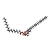 ChemComp-6OU: |
-Macromolecule #4: CHOLESTEROL
| Macromolecule | Name: CHOLESTEROL / type: ligand / ID: 4 / Number of copies: 2 / Formula: CLR |
|---|---|
| Molecular weight | Theoretical: 386.654 Da |
| Chemical component information |  ChemComp-CLR: |
-Macromolecule #5: [(2~{R})-1-[2-azanylethoxy(oxidanyl)phosphoryl]oxy-3-hexadecanoyl...
| Macromolecule | Name: [(2~{R})-1-[2-azanylethoxy(oxidanyl)phosphoryl]oxy-3-hexadecanoyloxy-propan-2-yl] (4~{Z},7~{Z},10~{Z},13~{Z},16~{Z},19~{Z})-docosa-4,7,10,13,16,19-hexaenoate type: ligand / ID: 5 / Number of copies: 2 / Formula: A1L1C |
|---|---|
| Molecular weight | Theoretical: 764.023 Da |
-Macromolecule #6: 1-palmitoyl-2-oleoyl-sn-glycero-3-phosphocholine
| Macromolecule | Name: 1-palmitoyl-2-oleoyl-sn-glycero-3-phosphocholine / type: ligand / ID: 6 / Number of copies: 2 / Formula: LBN |
|---|---|
| Molecular weight | Theoretical: 760.076 Da |
| Chemical component information | 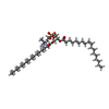 ChemComp-LBN: |
-Macromolecule #7: SODIUM ION
| Macromolecule | Name: SODIUM ION / type: ligand / ID: 7 / Number of copies: 2 |
|---|---|
| Molecular weight | Theoretical: 22.99 Da |
-Macromolecule #8: water
| Macromolecule | Name: water / type: ligand / ID: 8 / Number of copies: 2 / Formula: HOH |
|---|---|
| Molecular weight | Theoretical: 18.015 Da |
| Chemical component information |  ChemComp-HOH: |
-Experimental details
-Structure determination
| Method | cryo EM |
|---|---|
 Processing Processing | single particle reconstruction |
| Aggregation state | particle |
-Sample preparation
| Concentration | 9 mg/mL | |||||||||||||||
|---|---|---|---|---|---|---|---|---|---|---|---|---|---|---|---|---|
| Buffer | pH: 8 Component:
Details: 0.001 % LMNG, 150 mM NaCl, 0.2 mM TECP, 50 mM Tris HCl pH 8.0 | |||||||||||||||
| Grid | Model: Quantifoil R1.2/1.3 / Material: GOLD / Mesh: 300 / Support film - Material: CARBON / Support film - topology: HOLEY / Pretreatment - Type: GLOW DISCHARGE / Pretreatment - Time: 50 sec. / Pretreatment - Atmosphere: AIR / Pretreatment - Pressure: 0.026000000000000002 kPa | |||||||||||||||
| Vitrification | Cryogen name: ETHANE / Chamber humidity: 100 % / Chamber temperature: 277.15 K / Instrument: FEI VITROBOT MARK IV |
- Electron microscopy
| Microscope | FEI TITAN KRIOS |
|---|---|
| Image recording | Film or detector model: FEI FALCON IV (4k x 4k) / Average electron dose: 50.0 e/Å2 |
| Electron beam | Acceleration voltage: 300 kV / Electron source:  FIELD EMISSION GUN FIELD EMISSION GUN |
| Electron optics | Illumination mode: FLOOD BEAM / Imaging mode: BRIGHT FIELD / Nominal defocus max: 2.0 µm / Nominal defocus min: 1.5 µm |
| Experimental equipment |  Model: Titan Krios / Image courtesy: FEI Company |
+Image processing

-Atomic model buiding 1
| Refinement | Space: REAL |
|---|---|
| Output model | PDB-8z8z: |
