 Movie
Movie Controller

[English] 日本語
 Yorodumi
- EMDB-3982: Molecular Architecture of the Jumonji C histone demethylase KDM5B. -
+ Open data

- Basic information
| Entry | Database: EMDB / ID: EMD-3982 | |||||||||
|---|---|---|---|---|---|---|---|---|---|---|
| Title | Molecular Architecture of the Jumonji C histone demethylase KDM5B. | |||||||||
 Map data Map data | ||||||||||
 Sample Sample |
| |||||||||
| Biological species |  Homo sapiens (human) Homo sapiens (human) | |||||||||
| Method | single particle reconstruction / negative staining / Resolution: 27.0 Å | |||||||||
 Authors Authors | Aduri NA / Gajhede M | |||||||||
 Citation Citation | Journal: Sci Rep / Year: 2019 Title: Molecular architecture of the Jumonji C family histone demethylase KDM5B. Authors: Jerzy Dorosz / Line Hyltoft Kristensen / Nanda G Aduri / Osman Mirza / Rikke Lousen / Saskia Bucciarelli / Ved Mehta / Selene Sellés-Baiget / Sara Marie Øie Solbak / Anders Bach / Pablo ...Authors: Jerzy Dorosz / Line Hyltoft Kristensen / Nanda G Aduri / Osman Mirza / Rikke Lousen / Saskia Bucciarelli / Ved Mehta / Selene Sellés-Baiget / Sara Marie Øie Solbak / Anders Bach / Pablo Mesa / Pablo Alcon Hernandez / Guillermo Montoya / Tam T T N Nguyen / Kasper D Rand / Thomas Boesen / Michael Gajhede /  Abstract: The full length human histone 3 lysine 4 demethylase KDM5B (PLU-1/Jarid1B) has been studied using Hydrogen/Deuterium exchange mass spectrometry, homology modelling, sequence analysis, small angle X- ...The full length human histone 3 lysine 4 demethylase KDM5B (PLU-1/Jarid1B) has been studied using Hydrogen/Deuterium exchange mass spectrometry, homology modelling, sequence analysis, small angle X-ray scattering and electron microscopy. This first structure on an intact multi-domain Jumonji histone demethylase reveal that the so-called PLU region, in the central region of KDM5B, has a curved α-helical three-dimensional structure, that acts as a rigid linker between the catalytic core and a region comprising four α-helices, a loop comprising the PHD2 domain, two large intrinsically disordered loops and the PHD3 domain in close proximity. The dumbbell shaped and curved KDM5B architecture observed by electron microscopy is complementary to the nucleosome surface and has a striking overall similarity to that of the functionally related KDM1A/CoREST complex. This could suggest that there are similarities between the demethylation mechanisms employed by the two histone 3 lysine 4 demethylases at the molecular level. | |||||||||
| History |
|
- Structure visualization
| Movie |
 Movie viewer Movie viewer |
|---|---|
| Structure viewer | EM map: SurfViewMolmilJmol/JSmol |
| Supplemental images |
 UCSF Chimera
- Downloads & links
-EMDB archive
| Map data | emd_3982.map.gz | 17 MB | EMDB map data format | |
|---|---|---|---|---|
| Header (meta data) | emd-3982-v30.xmlemd-3982.xml | 9.7 KB 9.7 KB | Display Display | EMDB header |
| Images | 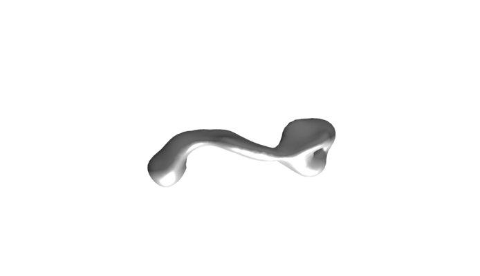 emd_3982.png emd_3982.png | 12.2 KB | ||
| Archive directory |  http://ftp.pdbj.org/pub/emdb/structures/EMD-3982ftp://ftp.pdbj.org/pub/emdb/structures/EMD-3982 http://ftp.pdbj.org/pub/emdb/structures/EMD-3982ftp://ftp.pdbj.org/pub/emdb/structures/EMD-3982 | HTTPS FTP |
-Related structure data
| Related structure data | C: citing same article ( |
|---|---|
| Similar structure data |


-Links
| EMDB pages | EMDB (EBI/PDBe) / EMDataResource |
|---|
-Map
| File | Download / File: emd_3982.map.gz / Format: CCP4 / Size: 22.2 MB / Type: IMAGE STORED AS FLOATING POINT NUMBER (4 BYTES) | ||||||||||||||||||||||||||||||||||||||||||||||||||||||||||||||||||||
|---|---|---|---|---|---|---|---|---|---|---|---|---|---|---|---|---|---|---|---|---|---|---|---|---|---|---|---|---|---|---|---|---|---|---|---|---|---|---|---|---|---|---|---|---|---|---|---|---|---|---|---|---|---|---|---|---|---|---|---|---|---|---|---|---|---|---|---|---|---|
| Projections & slices | Image control
Images are generated by Spider. | ||||||||||||||||||||||||||||||||||||||||||||||||||||||||||||||||||||
| Voxel size | X=Y=Z: 3.15 Å | ||||||||||||||||||||||||||||||||||||||||||||||||||||||||||||||||||||
| Density |
| ||||||||||||||||||||||||||||||||||||||||||||||||||||||||||||||||||||
| Symmetry | Space group: 1 | ||||||||||||||||||||||||||||||||||||||||||||||||||||||||||||||||||||
| Details | EMDB XML:
CCP4 map header:
| ||||||||||||||||||||||||||||||||||||||||||||||||||||||||||||||||||||
 Z (Sec.)
 Y (Row.)
 X (Col.)


-Supplemental data
- Sample components
-Entire : KDM5B
| Entire | Name: KDM5B |
|---|---|
| Components |
|
-Supramolecule #1: KDM5B
| Supramolecule | Name: KDM5B / type: organelle_or_cellular_component / ID: 1 / Parent: 0 / Macromolecule list: all |
|---|---|
| Source (natural) | Organism: Homo sapiens (human) |
| Recombinant expression | Organism:  Trichoplusia ni (cabbage looper) Trichoplusia ni (cabbage looper) |
-Macromolecule #1: KDM5B
| Macromolecule | Name: KDM5B / type: protein_or_peptide / ID: 1 / Enantiomer: LEVO |
|---|---|
| Sequence | String: MEAATTLHPG PRPALPLGGP GPLGEFLPPP ECPVFEPSWE EFADPFAFIH KIRPIAEQTG ICKVRPPPD WQPPFACDVD KLHFTPRIQR LNELEAQTRV KLNFLDQIAK YWELQGSTLK I PHVERKIL DLFQLNKLVA EEGGFAVVCK DRKWTKIATK MGFAPGKAVG ...String: MEAATTLHPG PRPALPLGGP GPLGEFLPPP ECPVFEPSWE EFADPFAFIH KIRPIAEQTG ICKVRPPPD WQPPFACDVD KLHFTPRIQR LNELEAQTRV KLNFLDQIAK YWELQGSTLK I PHVERKIL DLFQLNKLVA EEGGFAVVCK DRKWTKIATK MGFAPGKAVG SHIRGHYERI LN PYNLFLS GDSLRCLQKP NLTTDTKDKE YKPHDIPQRQ SVQPSETCPP ARRAKRMRAE AMN IKIEPE ETTEARTHNL RRRMGCPTPK CENEKEMKSS IKQEPIERKD YIVENEKEKP KSRS KKATN AVDLYVCLLC GSGNDEDRLL LCDGCDDSYH TFCLIPPLHD VPKGDWRCPK CLAQE CSKP QEAFGFEQAA RDYTLRTFGE MADAFKSDYF NMPVHMVPTE LVEKEFWRLV STIEED VTV EYGADIASKE FGSGFPVRDG KIKLSPEEEE YLDSGWNLNN MPVMEQSVLA HITADIC GM KLPWLYVGMC FSSFCWHIED HWSYSINYLH WGEPKTWYGV PGYAAEQLEN VMKKLAPE L FVSQPDLLHQ LVTIMNPNTL MTHEVPVYRT NQCAGEFVIT FPRAYHSGFN QGFNFAEAV NFCTVDWLPL GRQCVEHYRL LHRYCVFSHD EMICKMASKA DVLDVVVAST VQKDMAIMIE DEKALRETV RKLGVIDSER MDFELLPDDE RQCVKCKTTC FMSAISCSCK PGLLVCLHHV K ELCSCPPY KYKLRYRYTL DDLYPMMNAL KLRAESYNEW ALNVNEALEA KINKKKSLVS FK ALIEESE MKKFPDNDLL RHLRLVTQDA EKCASVAQQL LNGKRQTRYR SGGGKSQNQL TVN ELRQFV TQLYALPCVL SQTPLLKDLL NRVEDFQQHS QKLLSEETPS AAELQDLLDV SFEF DVELP QLAEMRIRLE QARWLEEVQQ ACLDPSSLTL DDMRRLIDLG VGLAPYSAVE KAMAR LQEL LTVSEHWDDK AKSLLKARPR HSLNSLATAV KEIEEIPAYL PNGAALKDSV QRARDW LQD VEGLQAGGRV PVLDTLIELV TRGRSIPVHL NSLPRLETLV AEVQAWKECA VNTFLTE NS PYSLLEVLCP RCDIGLLGLK RKQRKLKEPL PNGKKKSTKL ESLSDLERAL TESKETAS A MATLGEARLR EMEALQSLRL ANEGKLLSPL QDVDIKICLC QKAPAAPMIQ CELCRDAFH TSCVAVPSIS QGLRIWLCPH CRRSEKPPLE KILPLLASLQ RIRVRLPEGD ALRYMIERTV NWQHRAQQL LSSGNLKFVQ DRVGSGLLYS RWQASAGQVS DTNKVSQPPG TTSFSLPDDW D NRTSYLHS PFSTGRSCIP LHGVSPEVNE LLMEAQLLQV SLPEIQELYQ TLLAKPSPAQ QT DRSSPVR PSSEKNDCCR GKRDGINSLE RKLKRRLERE GLSSERWERV KKMRTPKKKK IKL SHPKDM NNFKLERERS YELVRSAETH SLPSDTSYSE QEDSEDEDAI CPAVSCLQPE GDEV DWVQC DGSCNQWFHQ VCVGVSPEMA EKEDYICVRC TVKDAPSRK |
-Experimental details
-Structure determination
| Method | negative staining |
|---|---|
 Processing Processing | single particle reconstruction |
| Aggregation state | particle |
-Sample preparation
| Buffer | pH: 7.7 |
|---|---|
| Staining | Type: NEGATIVE / Material: Uranyl formate |
- Electron microscopy
| Microscope | FEI TECNAI SPIRIT |
|---|---|
| Image recording | Film or detector model: TVIPS TEMCAM-F416 (4k x 4k) / Average electron dose: 10.0 e/Å2 |
| Electron beam | Acceleration voltage: 120 kV / Electron source: LAB6 |
| Electron optics | Illumination mode: SPOT SCAN / Imaging mode: DARK FIELD |
| Experimental equipment |  Model: Tecnai Spirit / Image courtesy: FEI Company |
-Image processing
| Final reconstruction | Resolution.type: BY AUTHOR / Resolution: 27.0 Å / Resolution method: FSC 0.143 CUT-OFF / Number images used: 9599 |
|---|---|
| Initial angle assignment | Type: NOT APPLICABLE |
| Final angle assignment | Type: NOT APPLICABLE |
