Movie
Movie Controller

[English] 日本語
 Yorodumi
- EMDB-39481: 2.08A Apoferritin Structure Solved Using an Indirect Scintillator... -
+ Open data

- Basic information
| Entry |  | |||||||||
|---|---|---|---|---|---|---|---|---|---|---|
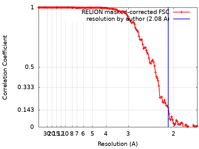
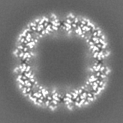
| Title | 2.08A Apoferritin Structure Solved Using an Indirect Scintillator-Coupled CMOS Detector at 300 kV | |||||||||
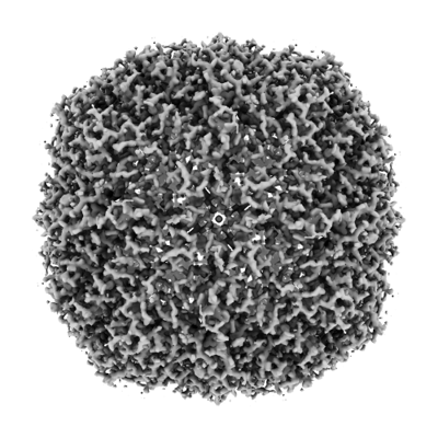
 Map data Map data | FSC-weighted, shapened and masked map by PostProcess. | |||||||||
 Sample Sample |
| |||||||||
 Keywords Keywords | apoferritin / ferritin / METAL BINDING PROTEIN | |||||||||
| Biological species |  | |||||||||
| Method | single particle reconstruction / cryo EM / Resolution: 2.08 Å | |||||||||
 Authors Authors | Aramaki S / Yoshida Y / Tanihara T / Oyama K / Otsuki K / Terada Y / Matsunaga N / Ohdo S / Mayanagi K | |||||||||
| Funding support | 1 items
| |||||||||
 Citation Citation | Journal: To Be Published Title: High-Resolution Single Particle Analysis Using an Indirect Scintillator-Coupled CMOS Detector at 300 kV Authors: Aramaki S / Yoshida Y / Tanihara T / Oyama K / Otsuki K / Terada Y / Matsunaga N / Ohdo S / Mayanagi K | |||||||||
| History |
|
- Structure visualization
| Supplemental images |
|---|
- Downloads & links
-EMDB archive
| Map data | emd_39481.map.gz | 13.9 MB |  EMDB map data format EMDB map data format | |
|---|---|---|---|---|
| Header (meta data) | emd-39481-v30.xmlemd-39481.xml | 15.7 KB 15.7 KB | Display Display | EMDB header |
| FSC (resolution estimation) | emd_39481_fsc.xml | 12.3 KB | Display | FSC data file |
| Images |  emd_39481.png emd_39481.png | 140 KB | ||
| Masks | emd_39481_msk_1.map | 22.2 MB | Mask map | |
| Filedesc metadata | emd-39481.cif.gz | 4.6 KB | ||
| Others | emd_39481_half_map_1.map.gzemd_39481_half_map_2.map.gz | 16.8 MB 16.8 MB | ||
| Archive directory |  http://ftp.pdbj.org/pub/emdb/structures/EMD-39481ftp://ftp.pdbj.org/pub/emdb/structures/EMD-39481 http://ftp.pdbj.org/pub/emdb/structures/EMD-39481ftp://ftp.pdbj.org/pub/emdb/structures/EMD-39481 | HTTPS FTP |
-Validation report
| Summary document | emd_39481_validation.pdf.gz | 859.1 KB | Display | EMDB validaton report |
|---|---|---|---|---|
| Full document | emd_39481_full_validation.pdf.gz | 858.6 KB | Display | |
| Data in XML | emd_39481_validation.xml.gz | 15.6 KB | Display | |
| Data in CIF | emd_39481_validation.cif.gz | 20.7 KB | Display | |
| Arichive directory | https://ftp.pdbj.org/pub/emdb/validation_reports/EMD-39481ftp://ftp.pdbj.org/pub/emdb/validation_reports/EMD-39481 | HTTPS FTP |
-Related structure data

-Links
| EMDB pages | EMDB (EBI/PDBe) / EMDataResource |
|---|
-Map
| File | Download / File: emd_39481.map.gz / Format: CCP4 / Size: 22.2 MB / Type: IMAGE STORED AS FLOATING POINT NUMBER (4 BYTES) | ||||||||||||||||||||
|---|---|---|---|---|---|---|---|---|---|---|---|---|---|---|---|---|---|---|---|---|---|
| Annotation | FSC-weighted, shapened and masked map by PostProcess. | ||||||||||||||||||||
| Voxel size | X=Y=Z: 0.8512 Å | ||||||||||||||||||||
| Density |
| ||||||||||||||||||||
| Symmetry | Space group: 1 | ||||||||||||||||||||
| Details | EMDB XML:
|
-Supplemental data
-Mask #1
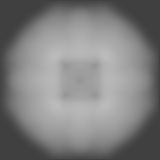
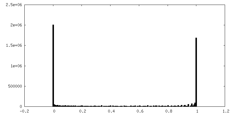
| File | emd_39481_msk_1.map | ||||||||||||
|---|---|---|---|---|---|---|---|---|---|---|---|---|---|
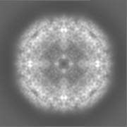
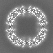
| Projections & Slices |
| ||||||||||||
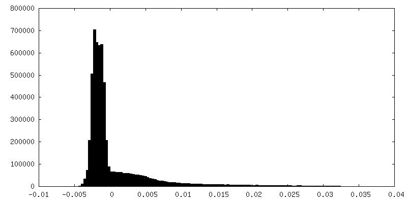
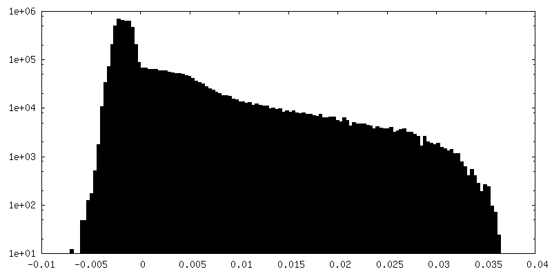
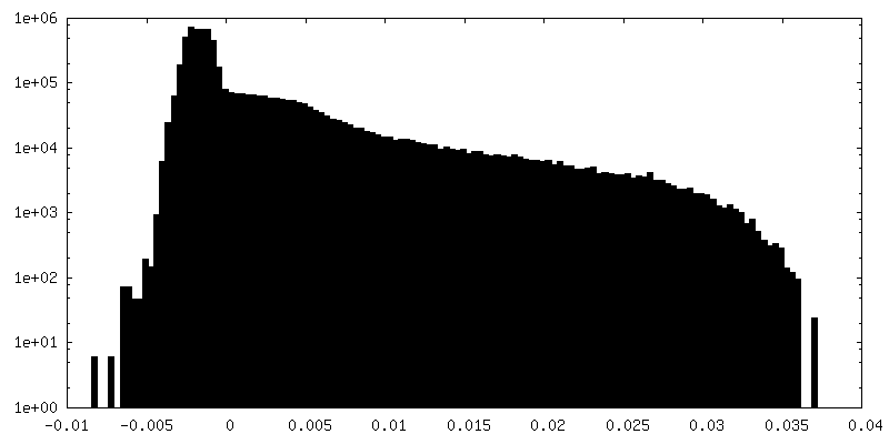
| Density Histograms |
 Z
Z Y
Y X
X


-Half map: #1
| File | emd_39481_half_map_1.map | ||||||||||||
|---|---|---|---|---|---|---|---|---|---|---|---|---|---|
| Projections & Slices |
| ||||||||||||
| Density Histograms |


-Half map: #2
| File | emd_39481_half_map_2.map | ||||||||||||
|---|---|---|---|---|---|---|---|---|---|---|---|---|---|
| Projections & Slices |
| ||||||||||||
| Density Histograms |


- Sample components
-Entire : apoferritin
| Entire | Name: apoferritin |
|---|---|
| Components |
|
-Supramolecule #1: apoferritin
| Supramolecule | Name: apoferritin / type: complex / ID: 1 / Parent: 0 |
|---|---|
| Source (natural) | Organism: |
| Molecular weight | Theoretical: 440 KDa |
-Experimental details
-Structure determination
| Method | cryo EM |
|---|---|
 Processing Processing | single particle reconstruction |
| Aggregation state | particle |
-Sample preparation
| Concentration | 11 mg/mL | ||||||||||||
|---|---|---|---|---|---|---|---|---|---|---|---|---|---|
| Buffer | pH: 7.5 Component:
| ||||||||||||
| Grid | Model: Quantifoil / Material: COPPER / Mesh: 200 / Support film - Material: CARBON / Support film - topology: HOLEY ARRAY / Pretreatment - Type: GLOW DISCHARGE / Pretreatment - Atmosphere: AIR | ||||||||||||
| Vitrification | Cryogen name: ETHANE / Chamber humidity: 100 % / Chamber temperature: 277.15 K / Instrument: FEI VITROBOT MARK IV |
- Electron microscopy
| Microscope | JEOL CRYO ARM 300 |
|---|---|
| Specialist optics | Phase plate: OTHER / Energy filter - Name: In-column Omega Filter / Energy filter - Slit width: 20 eV |
| Image recording | Film or detector model: OTHER / Digitization - Dimensions - Width: 4096 pixel / Digitization - Dimensions - Height: 4096 pixel / Number grids imaged: 1 / Number real images: 1893 / Average exposure time: 2.3 sec. / Average electron dose: 92.9 e/Å2 / Details: TVIPS TemCam-XF416 (4k x 4k) |
| Electron beam | Acceleration voltage: 300 kV / Electron source:  FIELD EMISSION GUN FIELD EMISSION GUN |
| Electron optics | C2 aperture diameter: 50.0 µm / Illumination mode: SPOT SCAN / Imaging mode: BRIGHT FIELD / Cs: 2.7 mm / Nominal defocus max: 1.5 µm / Nominal defocus min: 0.2 µm / Nominal magnification: 200000 |
| Sample stage | Specimen holder model: JEOL CRYOSPECPORTER / Cooling holder cryogen: NITROGEN |