 Movie
Movie Controller


+ Open data

- Basic information
| Entry |  | |||||||||
|---|---|---|---|---|---|---|---|---|---|---|
| Title | structure of phage T4 topoisomerase II central domain | |||||||||
 Map data Map data | T4 Topo II central domain map | |||||||||
 Sample Sample |
| |||||||||
 Keywords Keywords | topoisomerase II / ISOMERASE | |||||||||
| Function / homology |  Function and homology information Function and homology informationsister chromatid segregation / DNA topoisomerase type II (double strand cut, ATP-hydrolyzing) activity / DNA topoisomerase (ATP-hydrolysing) / DNA topological change / protein-containing complex / DNA binding / ATP binding Similarity search - Function | |||||||||
| Biological species |  Escherichia phage T4 (virus) Escherichia phage T4 (virus) | |||||||||
| Method | single particle reconstruction / cryo EM / Resolution: 3.62 Å | |||||||||
 Authors Authors | Chen YT / Xin YH / Xian RQ | |||||||||
| Funding support |  China, 1 items China, 1 items
| |||||||||
 Citation Citation | Journal: Nat Commun / Year: 2024 Title: Structural and functional insights into the T-even type bacteriophage topoisomerase II. Authors: Yuhui Xin / Runqi Xian / Yunge Yang / Jingyuan Cong / Zihe Rao / Xuemei Li / Yutao Chen / Abstract: T-even type bacteriophages are virulent phages commonly used as model organisms, playing a crucial role in understanding various biological processes. One such process involves the regulation of DNA ...T-even type bacteriophages are virulent phages commonly used as model organisms, playing a crucial role in understanding various biological processes. One such process involves the regulation of DNA topology during phage replication upon host infection, governed by type IIA DNA topoisomerases. In spite of various studies on prokaryotic and eukaryotic counterparts, viral topoisomerase II remains insufficiently understood, especially the unique domain composition of T4 phage. In this study, we determine the cryo-EM structures of topoisomerase II from T4 and T6 phages, including full-length structures of both apo and DNA-binding states which have never been determined before. Together with other conformational states, these structures provide an explicit blueprint of mechanisms of phage topoisomerase II. Particularly, the asymmetric dimeric interactions observed in cryo-EM structures of T6 phage topoisomerase II ATPase domain and central domain bound with DNA shed light on the asynchronous ATP usage and asynchronous cleavage of the G-segment DNA, respectively. The elucidation of phage topoisomerase II's structures and functions not only enhances our understanding of mechanisms and evolutionary parallels with prokaryotic and eukaryotic homologs but also highlights its potential as a model for developing type IIA topoisomerase inhibitors. | |||||||||
| History |
|
- Structure visualization
| Supplemental images |
|---|

- Downloads & links
-EMDB archive
| Map data | emd_39434.map.gz | 59.7 MB | EMDB map data format | |
|---|---|---|---|---|
| Header (meta data) | emd-39434-v30.xmlemd-39434.xml | 17.3 KB 17.3 KB | Display Display | EMDB header |
| Images |  emd_39434.png emd_39434.png | 104.1 KB | ||
| Filedesc metadata | emd-39434.cif.gz | 6.1 KB | ||
| Others | emd_39434_half_map_1.map.gzemd_39434_half_map_2.map.gz | 59.2 MB 59.2 MB | ||
| Archive directory |  http://ftp.pdbj.org/pub/emdb/structures/EMD-39434ftp://ftp.pdbj.org/pub/emdb/structures/EMD-39434 http://ftp.pdbj.org/pub/emdb/structures/EMD-39434ftp://ftp.pdbj.org/pub/emdb/structures/EMD-39434 | HTTPS FTP |
-Validation report
| Summary document | emd_39434_validation.pdf.gz | 775.5 KB | Display | EMDB validaton report |
|---|---|---|---|---|
| Full document | emd_39434_full_validation.pdf.gz | 775.1 KB | Display | |
| Data in XML | emd_39434_validation.xml.gz | 11.4 KB | Display | |
| Data in CIF | emd_39434_validation.cif.gz | 13.6 KB | Display | |
| Arichive directory | https://ftp.pdbj.org/pub/emdb/validation_reports/EMD-39434ftp://ftp.pdbj.org/pub/emdb/validation_reports/EMD-39434 | HTTPS FTP |
-Related structure data
| Related structure data |  8yo3MC  8yluC  8yo1C 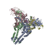 8yo4C 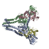 8yo5C 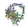 8yo7C 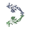 8yo9C 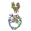 8yodC 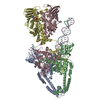 8yonC 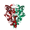 9imjC M: atomic model generated by this map C: citing same article ( |
|---|---|
| Similar structure data |
-Links
| EMDB pages | EMDB (EBI/PDBe) / EMDataResource |
|---|---|
| Related items in Molecule of the Month |


-Map
| File | Download / File: emd_39434.map.gz / Format: CCP4 / Size: 64 MB / Type: IMAGE STORED AS FLOATING POINT NUMBER (4 BYTES) | ||||||||||||||||||||||||||||||||||||
|---|---|---|---|---|---|---|---|---|---|---|---|---|---|---|---|---|---|---|---|---|---|---|---|---|---|---|---|---|---|---|---|---|---|---|---|---|---|
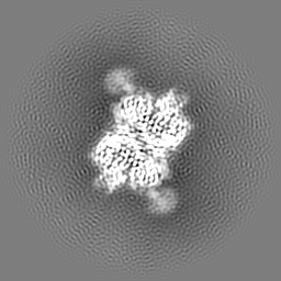
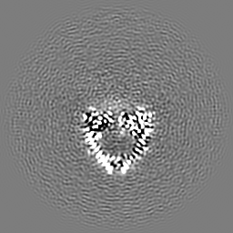
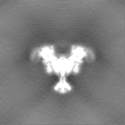
| Annotation | T4 Topo II central domain map | ||||||||||||||||||||||||||||||||||||
| Projections & slices | Image control
Images are generated by Spider. | ||||||||||||||||||||||||||||||||||||
| Voxel size | X=Y=Z: 1 Å | ||||||||||||||||||||||||||||||||||||
| Density |
| ||||||||||||||||||||||||||||||||||||
| Symmetry | Space group: 1 | ||||||||||||||||||||||||||||||||||||
| Details | EMDB XML:
|
 Z (Sec.)
 Y (Row.)
 X (Col.)


-Supplemental data
-Half map: T4 Topo II central domain half map
| File | emd_39434_half_map_1.map | ||||||||||||
|---|---|---|---|---|---|---|---|---|---|---|---|---|---|
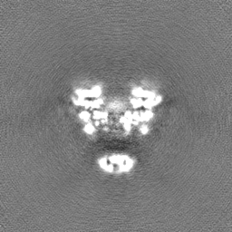
| Annotation | T4 Topo II central domain half map | ||||||||||||
| Projections & Slices |
| ||||||||||||
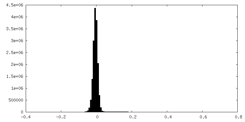
| Density Histograms |


-Half map: T4 Topo II central domain half map
| File | emd_39434_half_map_2.map | ||||||||||||
|---|---|---|---|---|---|---|---|---|---|---|---|---|---|
| Annotation | T4 Topo II central domain half map | ||||||||||||
| Projections & Slices |
| ||||||||||||
| Density Histograms |


- Sample components
-Entire : Phage T4 topoisomerase II central domian
| Entire | Name: Phage T4 topoisomerase II central domian |
|---|---|
| Components |
|
-Supramolecule #1: Phage T4 topoisomerase II central domian
| Supramolecule | Name: Phage T4 topoisomerase II central domian / type: complex / ID: 1 / Parent: 0 / Macromolecule list: all |
|---|---|
| Source (natural) | Organism: Escherichia phage T4 (virus) |
-Macromolecule #1: DNA topoisomerase medium subunit
| Macromolecule | Name: DNA topoisomerase medium subunit / type: protein_or_peptide / ID: 1 / Number of copies: 2 / Enantiomer: LEVO / EC number: DNA topoisomerase (ATP-hydrolysing) |
|---|---|
| Source (natural) | Organism: Escherichia phage T4 (virus) |
| Molecular weight | Theoretical: 50.570523 KDa |
| Recombinant expression | Organism:  |
| Sequence | String: MQLNNRDLKS IIDNEALAYA MYTVENRAIP NMIDGFKPVQ RFVIARALDL ARGNKDKFHK LASIAGGVAD LGYHHGENSA QDAGALMAN TWNNNFPLLD GQGNFGSRTV QKAAASRYIF ARVSKNFYNV YKDTEYAPVH QDKEHIPPAF YLPIIPTVLL N GVSGIATG ...String: MQLNNRDLKS IIDNEALAYA MYTVENRAIP NMIDGFKPVQ RFVIARALDL ARGNKDKFHK LASIAGGVAD LGYHHGENSA QDAGALMAN TWNNNFPLLD GQGNFGSRTV QKAAASRYIF ARVSKNFYNV YKDTEYAPVH QDKEHIPPAF YLPIIPTVLL N GVSGIATG YATYILPHSV SSVKKAVLQA LQGKKVTKPK VEFPEFRGEV VEIDGQYEIR GTYKFTSRTQ MHITEIPYKY DR ETYVSKI LDPLENKGFI TWDDACGEHG FGFKVKFRKE YSLSDNEEER HAKIMKDFGL IERRSQNITV INEKGKLQVY DNV VDLIKD FVEVRKTYVQ KRIDNKIKET ESAFRLAFAK AHFIKKVISG EIVVQGKTRK ELTEELSKID MYSSYVDKLV GMNI FHMTS DEAKKLAEEA KAKKEENEYW KTTDVVTEYT KDLEEIK UniProtKB: DNA topoisomerase medium subunit |
-Macromolecule #2: phage T4 topoisomerase II gp39-gp60 subunit
| Macromolecule | Name: phage T4 topoisomerase II gp39-gp60 subunit / type: protein_or_peptide / ID: 2 / Number of copies: 2 / Enantiomer: LEVO |
|---|---|
| Source (natural) | Organism: Escherichia phage T4 (virus) |
| Molecular weight | Theoretical: 77.524703 KDa |
| Recombinant expression | Organism: |
| Sequence | String: MIKNEIKILS DIEHIKKRSG MYIGSSANET HERFMFGKWE SVQYVPGLVK LIDEIIDNSV DEGIRTKFKF ANKINVTIKN NQVTVEDNG RGIPQAMVKT PTGEEIPGPV AAWTIPKAGG NFGDDKERVT GGMNGVGSSL TNIFSVMFVG ETGDGQNNIV V RCSNGMEN ...String: MIKNEIKILS DIEHIKKRSG MYIGSSANET HERFMFGKWE SVQYVPGLVK LIDEIIDNSV DEGIRTKFKF ANKINVTIKN NQVTVEDNG RGIPQAMVKT PTGEEIPGPV AAWTIPKAGG NFGDDKERVT GGMNGVGSSL TNIFSVMFVG ETGDGQNNIV V RCSNGMEN KSWEDIPGKW KGTRVTFIPD FMSFETNELS QVYLDITLDR LQTLAVVYPD IQFTFNGKKV QGNFKKYARQ YD EHAIVQE QENCSIAVGR SPDGFRQLTY VNNIHTKNGG HHIDCAMDDI CEDLIPQIKR KFKIDVTKAR VKECLTIVMF VRD MKNMRL IRQTKERLTS PFGEIRSHIQ LDAKKISRDI LNNEAILMPI IEAALARKLA AEKAAETKAA KKASKAKVHK HIKA NLCGK DADTTLFLTE GDSAIGYLID VRDKELHGGY PLRGKVLNSW GMSYADMLKN KELFDICAIT GLVLGEKAFE EKEDG EWFT FELNGDTIIV NENDEVQING KWITVGELRK NLMKFVKIDS SSVDMKKYKL QNNVRRSIKS SSMNYANVAI MTDADH DGL GSIYPSLLGF FSNWPELFEQ GRIRFVKTPV IIAQVGKKQE WFYTVAEYES AKDALPKHSI RYIKGLGSLE KSEYREM IQ NPVYDVVKLP ENWKELFEML MGDNADLRKE WMSQHHHHHH |
-Experimental details
-Structure determination
| Method | cryo EM |
|---|---|
 Processing Processing | single particle reconstruction |
| Aggregation state | particle |
-Sample preparation
| Buffer | pH: 6 |
|---|---|
| Vitrification | Cryogen name: ETHANE |
- Electron microscopy
| Microscope | FEI TALOS ARCTICA |
|---|---|
| Image recording | Film or detector model: GATAN K2 QUANTUM (4k x 4k) / Detector mode: COUNTING / Average electron dose: 60.0 e/Å2 |
| Electron beam | Acceleration voltage: 200 kV / Electron source:  FIELD EMISSION GUN FIELD EMISSION GUN |
| Electron optics | Illumination mode: FLOOD BEAM / Imaging mode: BRIGHT FIELD / Nominal defocus max: 2.5 µm / Nominal defocus min: 1.5 µm |
| Experimental equipment |  Model: Talos Arctica / Image courtesy: FEI Company |
