 Movie
Movie Controller

[English] 日本語
 Yorodumi
- PDB-8yo7: structure of phage T6 topoisomerase II central domain bound with ... -
+ Open data

- Basic information
| Entry | Database: PDB / ID: 8yo7 | ||||||
|---|---|---|---|---|---|---|---|
| Title | structure of phage T6 topoisomerase II central domain bound with DNA and m-AMSA | ||||||
 Components Components |
| ||||||
 Keywords Keywords | ISOMERASE / topoisomerase II | ||||||
| Function / homology |  Function and homology information Function and homology informationsister chromatid segregation / DNA topoisomerase type II (double strand cut, ATP-hydrolyzing) activity / DNA topoisomerase (ATP-hydrolysing) / DNA topological change / protein-containing complex / DNA binding / ATP binding / metal ion binding Similarity search - Function | ||||||
| Biological species |  Escherichia phage T4 (virus)Enterobacteria phage T6 (virus) Escherichia phage T4 (virus)Enterobacteria phage T6 (virus)DNA molecule (others) | ||||||
| Method | ELECTRON MICROSCOPY / single particle reconstruction / cryo EM / Resolution: 3.16 Å | ||||||
 Authors Authors | Chen, Y.T. / Xin, Y.H. / Xian, R.Q. | ||||||
| Funding support |  China, 1items China, 1items
| ||||||
 Citation Citation | Journal: Nat Commun / Year: 2024 Title: Structural and functional insights into the T-even type bacteriophage topoisomerase II. Authors: Yuhui Xin / Runqi Xian / Yunge Yang / Jingyuan Cong / Zihe Rao / Xuemei Li / Yutao Chen / Abstract: T-even type bacteriophages are virulent phages commonly used as model organisms, playing a crucial role in understanding various biological processes. One such process involves the regulation of DNA ...T-even type bacteriophages are virulent phages commonly used as model organisms, playing a crucial role in understanding various biological processes. One such process involves the regulation of DNA topology during phage replication upon host infection, governed by type IIA DNA topoisomerases. In spite of various studies on prokaryotic and eukaryotic counterparts, viral topoisomerase II remains insufficiently understood, especially the unique domain composition of T4 phage. In this study, we determine the cryo-EM structures of topoisomerase II from T4 and T6 phages, including full-length structures of both apo and DNA-binding states which have never been determined before. Together with other conformational states, these structures provide an explicit blueprint of mechanisms of phage topoisomerase II. Particularly, the asymmetric dimeric interactions observed in cryo-EM structures of T6 phage topoisomerase II ATPase domain and central domain bound with DNA shed light on the asynchronous ATP usage and asynchronous cleavage of the G-segment DNA, respectively. The elucidation of phage topoisomerase II's structures and functions not only enhances our understanding of mechanisms and evolutionary parallels with prokaryotic and eukaryotic homologs but also highlights its potential as a model for developing type IIA topoisomerase inhibitors. | ||||||
| History |
|
- Structure visualization
| Structure viewer | Molecule: MolmilJmol/JSmol |
|---|
- Downloads & links
-Download
| PDBx/mmCIF format | 8yo7.cif.gz | 304.2 KB | Display | PDBx/mmCIF format |
|---|---|---|---|---|
| PDB format | pdb8yo7.ent.gz | 235 KB | Display | PDB format |
| PDBx/mmJSON format | 8yo7.json.gz | Tree view | PDBx/mmJSON format | |
| Others |  Other downloads Other downloads |
-Validation report
| Arichive directory | https://data.pdbj.org/pub/pdb/validation_reports/yo/8yo7ftp://data.pdbj.org/pub/pdb/validation_reports/yo/8yo7 | HTTPS FTP |
|---|
-Related structure data
| Related structure data |  39437MC  8yluC  8yo1C  8yo3C 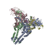 8yo4C 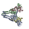 8yo5C 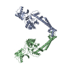 8yo9C 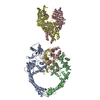 8yodC 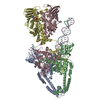 8yonC 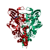 9imjC M: map data used to model this data C: citing same article ( |
|---|---|
| Similar structure data |
-Links
 PDBj
PDBj


- Assembly
| Deposited unit | 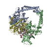
|
|---|---|
| 1 |
|
-Components
-DNA topoisomerase ... , 2 types, 4 molecules ABCD
| #1: Protein | Mass: 51951.973 Da / Num. of mol.: 2 Source method: isolated from a genetically manipulated source Source: (gene. exp.) Escherichia phage T4 (virus) / Gene: 52 / Production host:  References: UniProt: P07065, DNA topoisomerase (ATP-hydrolysing) #2: Protein | Mass: 69237.289 Da / Num. of mol.: 2 Source method: isolated from a genetically manipulated source Source: (gene. exp.) Enterobacteria phage T6 (virus) / Gene: EcT6_00003 / Production host: References: UniProt: A0A346FJ89, DNA topoisomerase (ATP-hydrolysing) |
|---|
-DNA chain , 2 types, 4 molecules EYFX
| #3: DNA chain | Mass: 16058.391 Da / Num. of mol.: 2 / Source method: obtained synthetically / Source: (synth.) DNA molecule (others) #4: DNA chain | Mass: 15965.361 Da / Num. of mol.: 2 / Source method: obtained synthetically / Source: (synth.) DNA molecule (others) |
|---|
-Non-polymers , 3 types, 6 molecules 

| #5: Chemical |  Mass: 24.305 Da / Num. of mol.: 2 / Source method: obtained synthetically / Formula: Mg / Feature type: SUBJECT OF INVESTIGATION Mass: 24.305 Da / Num. of mol.: 2 / Source method: obtained synthetically / Formula: Mg / Feature type: SUBJECT OF INVESTIGATION#6: Chemical |  Mass: 393.459 Da / Num. of mol.: 2 / Source method: obtained synthetically / Formula: C21H19N3O3S / Feature type: SUBJECT OF INVESTIGATION / Comment: antineoplastic*YM Mass: 393.459 Da / Num. of mol.: 2 / Source method: obtained synthetically / Formula: C21H19N3O3S / Feature type: SUBJECT OF INVESTIGATION / Comment: antineoplastic*YM#7: Water | ChemComp-HOH / | Mass: 18.015 Da / Num. of mol.: 2 / Source method: isolated from a natural source / Formula: H2O |
|---|
-Details
| Has ligand of interest | Y |
|---|---|
| Has protein modification | N |
-Experimental details
-Experiment
| Experiment | Method: ELECTRON MICROSCOPY |
|---|---|
| EM experiment | Aggregation state: PARTICLE / 3D reconstruction method: single particle reconstruction |
- Sample preparation
| Component | Name: Phage T6 topoisomerase II central domain bound with DNA and m-AMSA Type: COMPLEX / Entity ID: #1-#4 / Source: RECOMBINANT |
|---|---|
| Source (natural) | Organism: Enterobacteria phage T6 (virus) |
| Source (recombinant) | Organism: |
| Buffer solution | pH: 6 |
| Specimen | Embedding applied: NO / Shadowing applied: NO / Staining applied: NO / Vitrification applied: YES |
| Vitrification | Cryogen name: ETHANE |
- Electron microscopy imaging
| Experimental equipment |  Model: Titan Krios / Image courtesy: FEI Company |
|---|---|
| Microscopy | Model: FEI TITAN KRIOS |
| Electron gun | Electron source:  FIELD EMISSION GUN / Accelerating voltage: 300 kV / Illumination mode: FLOOD BEAM FIELD EMISSION GUN / Accelerating voltage: 300 kV / Illumination mode: FLOOD BEAM |
| Electron lens | Mode: BRIGHT FIELD / Nominal defocus max: 2500 nm / Nominal defocus min: 1500 nm |
| Image recording | Electron dose: 60 e/Å2 / Detector mode: COUNTING / Film or detector model: GATAN K2 QUANTUM (4k x 4k) |
- Processing
| CTF correction | Type: PHASE FLIPPING ONLY |
|---|---|
| 3D reconstruction | Resolution: 3.16 Å / Resolution method: FSC 0.143 CUT-OFF / Num. of particles: 459464 / Symmetry type: POINT |
