Ministry of Education, Culture, Sports, Science and Technology (Japan)
JP17H05825
Japan
Ministry of Education, Culture, Sports, Science and Technology (Japan)
JP19H04845
Japan
Japan Society for the Promotion of Science (JSPS)
20H03078
Japan
Japan Agency for Medical Research and Development (AMED)
JP18am0101072
Japan
Citation
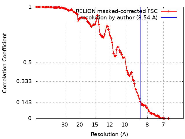
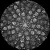
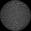
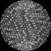
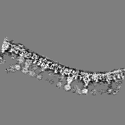
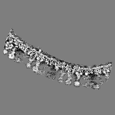
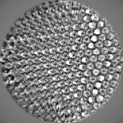
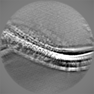
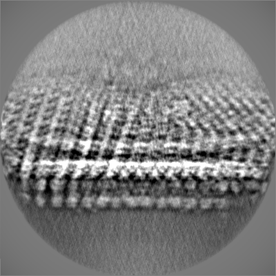
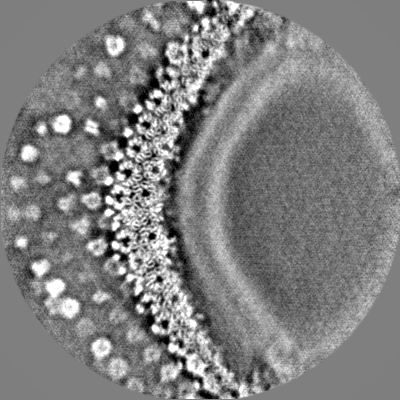
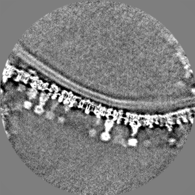
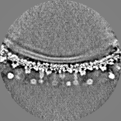
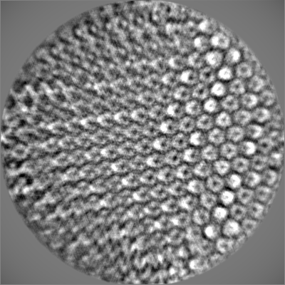
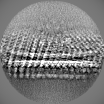
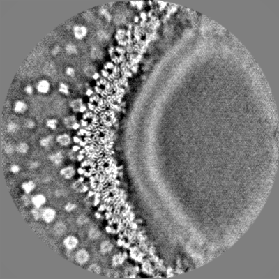
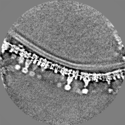
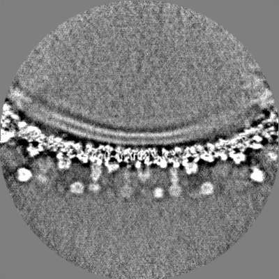
Journal: J Virol / Year: 2024 Title: Subnanometer structure of medusavirus capsid during maturation using cryo-electron microscopy. Authors: Ryoto Watanabe / Chihong Song / Masaharu Takemura / Kazuyoshi Murata / Abstract: Medusavirus is a giant virus classified into an independent family of . Amoebae infected with medusavirus release immature particles in addition to virions. These particles were suggested to exhibit ...Medusavirus is a giant virus classified into an independent family of . Amoebae infected with medusavirus release immature particles in addition to virions. These particles were suggested to exhibit the maturation process of this virus, but the structure of these capsids during maturation remains unknown. Here, we apply a block-based reconstruction method in cryo-electron microscopy (cryo-EM) single particle analysis to these viral capsids, extending the resolution to 7-10 Å. The maps reveal a novel network composed of minor capsid proteins (mCPs) supporting major capsid proteins (MCPs). A predicted molecular model of the MCP fitted into the cryo-EM maps clarified the boundaries between the MCP and the underlining mCPs, as well as between the MCP and the outer spikes, and identified molecular interactions between the MCP and these components. Several structural changes of the mCPs under the fivefold vertices of the immature particles were observed, depending on the presence or absence of the underlying internal membrane. In addition, the lower part of the penton proteins on the fivefold vertices was also missing in mature virions. These dynamic conformational changes of mCPs indicate an important function in the maturation process of medusavirus.IMPORTANCEThe structural changes of giant virus capsids during maturation have not thus far been well clarified. Medusavirus is a unique giant virus in which infected amoebae release immature particles in addition to mature virus particles. In this study, we used cryo-electron microscopy to investigate immature and mature medusavirus particles and elucidate the structural changes of the viral capsid during the maturation process. In DNA-empty particles, the conformation of the minor capsid proteins changed dynamically depending on the presence or absence of the underlying internal membranes. In DNA-full particles, the lower part of the penton proteins was lost. This is the first report of structural changes of the viral capsid during the maturation process of giant viruses.
In the structure databanks used in Yorodumi, some data are registered as the other names, "COVID-19 virus" and "2019-nCoV". Here are the details of the virus and the list of structure data.
Jan 31, 2019. EMDB accession codes are about to change! (news from PDBe EMDB page)
EMDB accession codes are about to change! (news from PDBe EMDB page)
The allocation of 4 digits for EMDB accession codes will soon come to an end. Whilst these codes will remain in use, new EMDB accession codes will include an additional digit and will expand incrementally as the available range of codes is exhausted. The current 4-digit format prefixed with “EMD-” (i.e. EMD-XXXX) will advance to a 5-digit format (i.e. EMD-XXXXX), and so on. It is currently estimated that the 4-digit codes will be depleted around Spring 2019, at which point the 5-digit format will come into force.
The EM Navigator/Yorodumi systems omit the EMD- prefix.
Related info.:Q: What is EMD? / ID/Accession-code notation in Yorodumi/EM Navigator
Yorodumi is a browser for structure data from EMDB, PDB, SASBDB, etc.
This page is also the successor to EM Navigator detail page, and also detail information page/front-end page for Omokage search.
The word "yorodu" (or yorozu) is an old Japanese word meaning "ten thousand". "mi" (miru) is to see.
Related info.:EMDB / PDB / SASBDB / Comparison of 3 databanks / Yorodumi Search / Aug 31, 2016. New EM Navigator & Yorodumi / Yorodumi Papers / Jmol/JSmol / Function and homology information / Changes in new EM Navigator and Yorodumi
 Movie
Movie Controller


 Open data
 Basic information
 Map data
 Sample
 Keywords
 Acanthamoeba castellanii medusavirus
 Authors
 Japan, 4 items
  Citation
 Structure visualization
 Downloads & links
 EMDB map data format
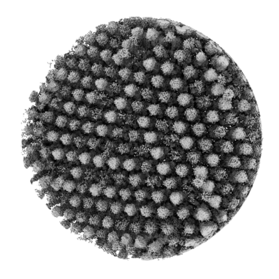 emd_39295.png
 http://ftp.pdbj.org/pub/emdb/structures/EMD-39295


 Z (Sec.)
 Y (Row.)
 X (Col.)


 Sample components
 Acanthamoeba castellanii (eukaryote)
 Processing
 Electron microscopy
 FIELD EMISSION GUN