 Movie
Movie Controller
 Structure viewers
 About Yorodumi Papers


+Search query
-Structure paper
| Title | Subnanometer structure of medusavirus capsid during maturation using cryo-electron microscopy. |
|---|---|
| Journal, issue, pages | J Virol, Vol. 98, Issue 9, Page e0043624, Year 2024 |
| Publish date | Sep 17, 2024 |
 Authors Authors | Ryoto Watanabe / Chihong Song / Masaharu Takemura / Kazuyoshi Murata /  |
| PubMed Abstract | Medusavirus is a giant virus classified into an independent family of . Amoebae infected with medusavirus release immature particles in addition to virions. These particles were suggested to exhibit ...Medusavirus is a giant virus classified into an independent family of . Amoebae infected with medusavirus release immature particles in addition to virions. These particles were suggested to exhibit the maturation process of this virus, but the structure of these capsids during maturation remains unknown. Here, we apply a block-based reconstruction method in cryo-electron microscopy (cryo-EM) single particle analysis to these viral capsids, extending the resolution to 7-10 Å. The maps reveal a novel network composed of minor capsid proteins (mCPs) supporting major capsid proteins (MCPs). A predicted molecular model of the MCP fitted into the cryo-EM maps clarified the boundaries between the MCP and the underlining mCPs, as well as between the MCP and the outer spikes, and identified molecular interactions between the MCP and these components. Several structural changes of the mCPs under the fivefold vertices of the immature particles were observed, depending on the presence or absence of the underlying internal membrane. In addition, the lower part of the penton proteins on the fivefold vertices was also missing in mature virions. These dynamic conformational changes of mCPs indicate an important function in the maturation process of medusavirus.IMPORTANCEThe structural changes of giant virus capsids during maturation have not thus far been well clarified. Medusavirus is a unique giant virus in which infected amoebae release immature particles in addition to mature virus particles. In this study, we used cryo-electron microscopy to investigate immature and mature medusavirus particles and elucidate the structural changes of the viral capsid during the maturation process. In DNA-empty particles, the conformation of the minor capsid proteins changed dynamically depending on the presence or absence of the underlying internal membranes. In DNA-full particles, the lower part of the penton proteins was lost. This is the first report of structural changes of the viral capsid during the maturation process of giant viruses. |
 External links External links | J Virol / PubMed:39194243 / PubMed Central |
| Methods | EM (single particle) |
| Resolution | 7.32 - 9.93 Å |
| Structure data | 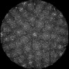 EMDB-39292: 5-fold block of DNA-Full medusavirus capsid 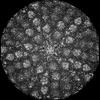 EMDB-39293: 5-fold block of DNA-Empty medusavirus capsid with internal membrane  EMDB-39294: 5-fold block of DNA-Empty medusavirus capsid without internal membrane 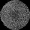 EMDB-39295: 3-fold block of DNA-Full medusavirus capsid  EMDB-39296: 3-fold block of DNA-Empty medusavirus capsid 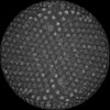 EMDB-39297: 2-fold block of DNA-Full medusavirus capsid 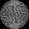 EMDB-39298: 2-fold block of DNA-Empty medusavirus capsid |
