 Movie
Movie Controller


+ Open data

- Basic information
| Entry | Database: EMDB / ID: EMD-3917 | |||||||||
|---|---|---|---|---|---|---|---|---|---|---|
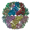
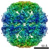
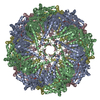
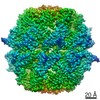
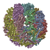
| Title | Rat TRiC structure | |||||||||
 Map data Map data | Rat TRiC | |||||||||
 Sample Sample |
| |||||||||
| Biological species |  | |||||||||
| Method | subtomogram averaging / cryo EM / Resolution: 17.1 Å | |||||||||
 Authors Authors | Guo Q / Lehmer C / Martinez-Sanchez A / Rudack T / Beck F / Hartmann H / Hipp MS / Hartl FU / Edbauer D / Baumeister W / Fernandez-Busnadiego R | |||||||||
 Citation Citation | Journal: Cell / Year: 2018 Title: In Situ Structure of Neuronal C9orf72 Poly-GA Aggregates Reveals Proteasome Recruitment. Authors: Qiang Guo / Carina Lehmer / Antonio Martínez-Sánchez / Till Rudack / Florian Beck / Hannelore Hartmann / Manuela Pérez-Berlanga / Frédéric Frottin / Mark S Hipp / F Ulrich Hartl / ...Authors: Qiang Guo / Carina Lehmer / Antonio Martínez-Sánchez / Till Rudack / Florian Beck / Hannelore Hartmann / Manuela Pérez-Berlanga / Frédéric Frottin / Mark S Hipp / F Ulrich Hartl / Dieter Edbauer / Wolfgang Baumeister / Rubén Fernández-Busnadiego /   Abstract: Protein aggregation and dysfunction of the ubiquitin-proteasome system are hallmarks of many neurodegenerative diseases. Here, we address the elusive link between these phenomena by employing cryo- ...Protein aggregation and dysfunction of the ubiquitin-proteasome system are hallmarks of many neurodegenerative diseases. Here, we address the elusive link between these phenomena by employing cryo-electron tomography to dissect the molecular architecture of protein aggregates within intact neurons at high resolution. We focus on the poly-Gly-Ala (poly-GA) aggregates resulting from aberrant translation of an expanded GGGGCC repeat in C9orf72, the most common genetic cause of amyotrophic lateral sclerosis and frontotemporal dementia. We find that poly-GA aggregates consist of densely packed twisted ribbons that recruit numerous 26S proteasome complexes, while other macromolecules are largely excluded. Proximity to poly-GA ribbons stabilizes a transient substrate-processing conformation of the 26S proteasome, suggesting stalled degradation. Thus, poly-GA aggregates may compromise neuronal proteostasis by driving the accumulation and functional impairment of a large fraction of cellular proteasomes. | |||||||||
| History |
|
- Structure visualization
| Movie |
 Movie viewer Movie viewer |
|---|---|
| Structure viewer | EM map: SurfViewMolmilJmol/JSmol |
| Supplemental images |

- Downloads & links
-EMDB archive
| Map data | emd_3917.map.gz | 758.7 KB | EMDB map data format | |
|---|---|---|---|---|
| Header (meta data) | emd-3917-v30.xmlemd-3917.xml | 16.6 KB 16.6 KB | Display Display | EMDB header |
| Images | 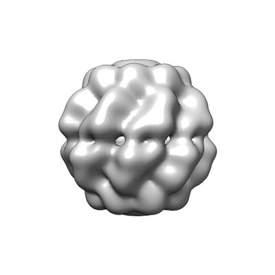 emd_3917.png emd_3917.png | 34.3 KB | ||
| Archive directory |  http://ftp.pdbj.org/pub/emdb/structures/EMD-3917ftp://ftp.pdbj.org/pub/emdb/structures/EMD-3917 http://ftp.pdbj.org/pub/emdb/structures/EMD-3917ftp://ftp.pdbj.org/pub/emdb/structures/EMD-3917 | HTTPS FTP |
-Related structure data
| Related structure data | 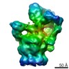 3913C 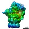 3914C 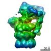 3915C 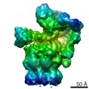 3916C  4191C 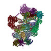 6epcC 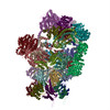 6epdC 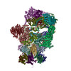 6epeC 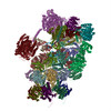 6epfC C: citing same article ( |
|---|---|
| Similar structure data |


-Links
| EMDB pages | EMDB (EBI/PDBe) / EMDataResource |
|---|
-Map
| File | Download / File: emd_3917.map.gz / Format: CCP4 / Size: 844.7 KB / Type: IMAGE STORED AS FLOATING POINT NUMBER (4 BYTES) | ||||||||||||||||||||||||||||||||||||||||||||||||||||||||||||
|---|---|---|---|---|---|---|---|---|---|---|---|---|---|---|---|---|---|---|---|---|---|---|---|---|---|---|---|---|---|---|---|---|---|---|---|---|---|---|---|---|---|---|---|---|---|---|---|---|---|---|---|---|---|---|---|---|---|---|---|---|---|
| Annotation | Rat TRiC | ||||||||||||||||||||||||||||||||||||||||||||||||||||||||||||
| Projections & slices | Image control
Images are generated by Spider. | ||||||||||||||||||||||||||||||||||||||||||||||||||||||||||||
| Voxel size | X=Y=Z: 3.42 Å | ||||||||||||||||||||||||||||||||||||||||||||||||||||||||||||
| Density |
| ||||||||||||||||||||||||||||||||||||||||||||||||||||||||||||
| Symmetry | Space group: 1 | ||||||||||||||||||||||||||||||||||||||||||||||||||||||||||||
| Details | EMDB XML:
CCP4 map header:
| ||||||||||||||||||||||||||||||||||||||||||||||||||||||||||||
 Z (Sec.)
 Y (Row.)
 X (Col.)


-Supplemental data
- Sample components
-Entire : Rat TRiC structure
| Entire | Name: Rat TRiC structure |
|---|---|
| Components |
|
-Supramolecule #1: Rat TRiC structure
| Supramolecule | Name: Rat TRiC structure / type: complex / ID: 1 / Parent: 0 / Macromolecule list: #1-#32 Details: in situ TRiC structure generated by subtomogram averaging |
|---|---|
| Source (natural) | Organism: |
-Experimental details
-Structure determination
| Method | cryo EM |
|---|---|
 Processing Processing | subtomogram averaging |
| Aggregation state | cell |
-Sample preparation
| Buffer | pH: 7 |
|---|---|
| Grid | Model: Quantifoil R2/1 / Material: GOLD / Mesh: 200 / Support film - Material: CARBON / Support film - topology: HOLEY / Pretreatment - Type: GLOW DISCHARGE / Pretreatment - Atmosphere: AIR / Details: The grid was coated with C prior to use |
| Vitrification | Cryogen name: ETHANE-PROPANE / Chamber humidity: 100 % / Chamber temperature: 298 K / Instrument: FEI VITROBOT MARK IV |
| Details | FIB milled rat primary neurons |
- Electron microscopy
| Microscope | FEI TITAN KRIOS |
|---|---|
| Image recording | Film or detector model: GATAN K2 SUMMIT (4k x 4k) / Detector mode: COUNTING / Digitization - Dimensions - Width: 3838 pixel / Digitization - Dimensions - Height: 3710 pixel / Average exposure time: 2.4 sec. / Average electron dose: 1.8 e/Å2 |
| Electron beam | Acceleration voltage: 300 kV / Electron source:  FIELD EMISSION GUN FIELD EMISSION GUN |
| Electron optics | C2 aperture diameter: 70.0 µm / Illumination mode: FLOOD BEAM / Imaging mode: BRIGHT FIELD / Cs: 2.7 mm / Nominal defocus max: 7.0 µm / Nominal defocus min: 5.0 µm / Nominal magnification: 42000 |
| Sample stage | Specimen holder model: FEI TITAN KRIOS AUTOGRID HOLDER / Cooling holder cryogen: NITROGEN |
| Experimental equipment |  Model: Titan Krios / Image courtesy: FEI Company |
+Image processing
-Atomic model buiding 1
| Refinement | Protocol: OTHER |
|---|
