 Movie
Movie Controller

[English] 日本語
 Yorodumi
- EMDB-39005: Focused refinement map of U5 snRNP of the human minor pre-B complex -
+ Open data

- Basic information
| Entry |  | ||||||||||||
|---|---|---|---|---|---|---|---|---|---|---|---|---|---|
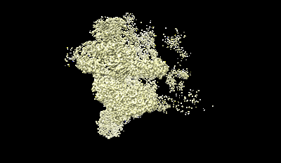
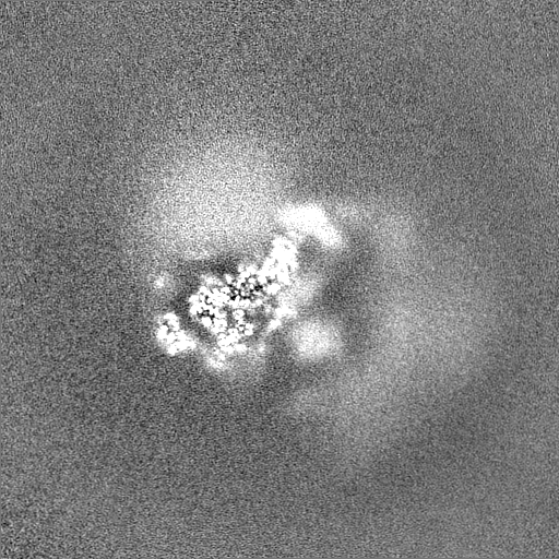
| Title | Focused refinement map of U5 snRNP of the human minor pre-B complex | ||||||||||||
 Map data Map data | |||||||||||||
 Sample Sample |
| ||||||||||||
 Keywords Keywords | spliceosome / minor spliceosome / U11 snRNP / U12 snRNP / U4atac / U6atac / CENATAC / TXNL4B / U11 snRNA / minor tri-snRNP / U12-type intron / SPLICING | ||||||||||||
| Biological species |  Homo sapiens (human) Homo sapiens (human) | ||||||||||||
| Method | single particle reconstruction / cryo EM / Resolution: 3.13 Å | ||||||||||||
 Authors Authors | Bai R / Yuan M / Zhang P / Luo T / Shi Y / Wan R | ||||||||||||
| Funding support |  China, 3 items China, 3 items
| ||||||||||||
 Citation Citation | Journal: Science / Year: 2024 Title: Structural basis of U12-type intron engagement by the fully assembled human minor spliceosome. Authors: Rui Bai / Meng Yuan / Pu Zhang / Ting Luo / Yigong Shi / Ruixue Wan / Abstract: The minor spliceosome, which is responsible for the splicing of U12-type introns, comprises five small nuclear RNAs (snRNAs), of which only one is shared with the major spliceosome. In this work, we ...The minor spliceosome, which is responsible for the splicing of U12-type introns, comprises five small nuclear RNAs (snRNAs), of which only one is shared with the major spliceosome. In this work, we report the 3.3-angstrom cryo-electron microscopy structure of the fully assembled human minor spliceosome pre-B complex. The atomic model includes U11 small nuclear ribonucleoprotein (snRNP), U12 snRNP, and U4atac/U6atac.U5 tri-snRNP. U11 snRNA is recognized by five U11-specific proteins (20K, 25K, 35K, 48K, and 59K) and the heptameric Sm ring. The 3' half of the 5'-splice site forms a duplex with U11 snRNA; the 5' half is recognized by U11-35K, U11-48K, and U11 snRNA. Two proteins, CENATAC and DIM2/TXNL4B, specifically associate with the minor tri-snRNP. A structural analysis uncovered how two conformationally similar tri-snRNPs are differentiated by the minor and major prespliceosomes for assembly. | ||||||||||||
| History |
|
- Structure visualization
| Supplemental images |
|---|
- Downloads & links
-EMDB archive
| Map data | emd_39005.map.gz | 481.6 MB |  EMDB map data format EMDB map data format | |
|---|---|---|---|---|
| Header (meta data) | emd-39005-v30.xmlemd-39005.xml | 17.4 KB 17.4 KB | Display Display | EMDB header |
| Images |  emd_39005.png emd_39005.png | 48.1 KB | ||
| Filedesc metadata | emd-39005.cif.gz | 4 KB | ||
| Others | emd_39005_half_map_1.map.gzemd_39005_half_map_2.map.gz | 472.9 MB 472.9 MB | ||
| Archive directory |  http://ftp.pdbj.org/pub/emdb/structures/EMD-39005ftp://ftp.pdbj.org/pub/emdb/structures/EMD-39005 http://ftp.pdbj.org/pub/emdb/structures/EMD-39005ftp://ftp.pdbj.org/pub/emdb/structures/EMD-39005 | HTTPS FTP |
-Related structure data


-Links
| EMDB pages | EMDB (EBI/PDBe) / EMDataResource |
|---|
-Map
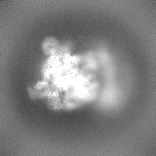
| File | Download / File: emd_39005.map.gz / Format: CCP4 / Size: 512 MB / Type: IMAGE STORED AS FLOATING POINT NUMBER (4 BYTES) | ||||||||||||||||||||||||||||||||||||
|---|---|---|---|---|---|---|---|---|---|---|---|---|---|---|---|---|---|---|---|---|---|---|---|---|---|---|---|---|---|---|---|---|---|---|---|---|---|
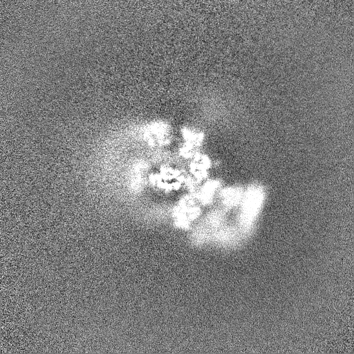
| Projections & slices | Image control
Images are generated by Spider. | ||||||||||||||||||||||||||||||||||||
| Voxel size | X=Y=Z: 1.0742 Å | ||||||||||||||||||||||||||||||||||||
| Density |
| ||||||||||||||||||||||||||||||||||||
| Symmetry | Space group: 1 | ||||||||||||||||||||||||||||||||||||
| Details | EMDB XML:
|
 Z (Sec.)
 Y (Row.)
 X (Col.)


-Supplemental data
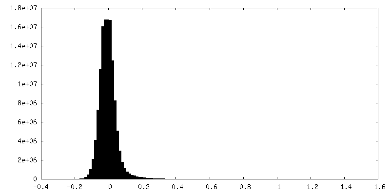
-Half map: #1
| File | emd_39005_half_map_1.map | ||||||||||||
|---|---|---|---|---|---|---|---|---|---|---|---|---|---|
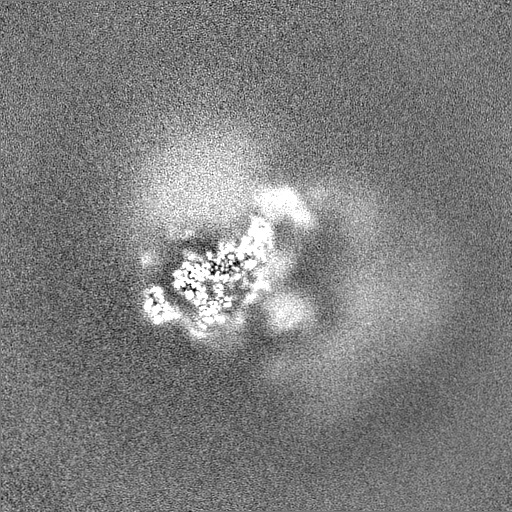
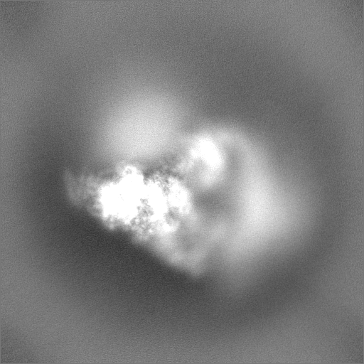
| Projections & Slices |
| ||||||||||||
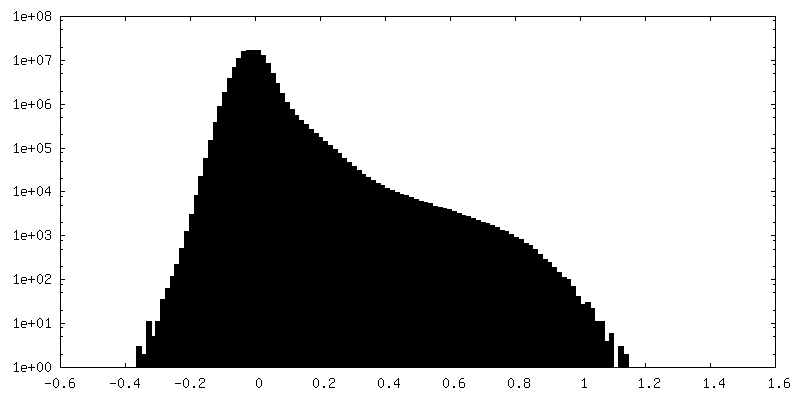
| Density Histograms |


-Half map: #2
| File | emd_39005_half_map_2.map | ||||||||||||
|---|---|---|---|---|---|---|---|---|---|---|---|---|---|
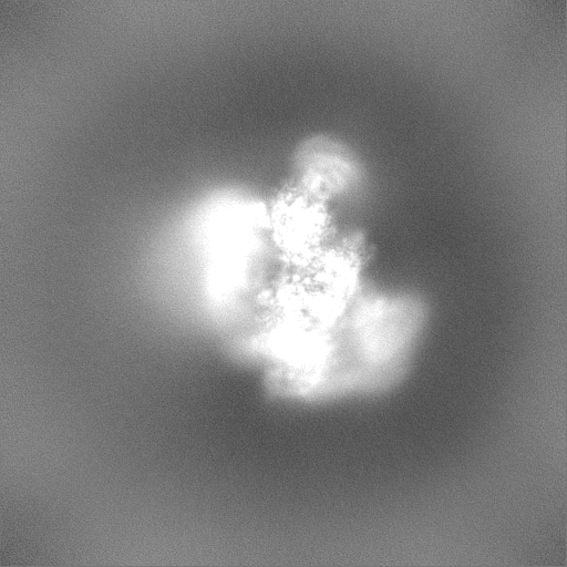
| Projections & Slices |
| ||||||||||||
| Density Histograms |


- Sample components
-Entire : human minor pre-B complex
| Entire | Name: human minor pre-B complex |
|---|---|
| Components |
|
-Supramolecule #1: human minor pre-B complex
| Supramolecule | Name: human minor pre-B complex / type: complex / ID: 1 / Parent: 0 / Macromolecule list: #1-#32 |
|---|---|
| Source (natural) | Organism: Homo sapiens (human) |
| Molecular weight | Theoretical: 1.8 MDa |
-Experimental details
-Structure determination
| Method | cryo EM |
|---|---|
 Processing Processing | single particle reconstruction |
| Aggregation state | particle |
-Sample preparation
| Concentration | 0.2 mg/mL |
|---|---|
| Buffer | pH: 7.9 |
| Grid | Model: Quantifoil R1.2/1.3 / Material: GOLD / Mesh: 300 / Support film - Material: CARBON / Support film - topology: CONTINUOUS / Pretreatment - Type: PLASMA CLEANING |
| Vitrification | Cryogen name: NITROGEN |
- Electron microscopy
| Microscope | FEI TITAN KRIOS |
|---|---|
| Image recording | Film or detector model: GATAN K3 (6k x 4k) / Average electron dose: 50.0 e/Å2 |
| Electron beam | Acceleration voltage: 300 kV / Electron source:  FIELD EMISSION GUN FIELD EMISSION GUN |
| Electron optics | Illumination mode: FLOOD BEAM / Imaging mode: BRIGHT FIELD / Nominal defocus max: 3.0 µm / Nominal defocus min: 0.5 µm / Nominal magnification: 81000 |
| Sample stage | Cooling holder cryogen: NITROGEN |
| Experimental equipment |  Model: Titan Krios / Image courtesy: FEI Company |
-Image processing
| Startup model | Type of model: INSILICO MODEL |
|---|---|
| Final reconstruction | Resolution.type: BY AUTHOR / Resolution: 3.13 Å / Resolution method: FSC 0.143 CUT-OFF / Number images used: 388888 |
| Initial angle assignment | Type: MAXIMUM LIKELIHOOD |
| Final angle assignment | Type: MAXIMUM LIKELIHOOD |
