 Movie
Movie Controller


+ Open data

- Basic information
| Entry | 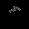 | |||||||||
|---|---|---|---|---|---|---|---|---|---|---|
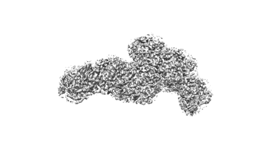
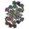
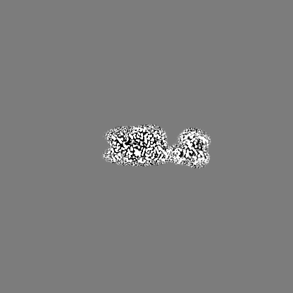
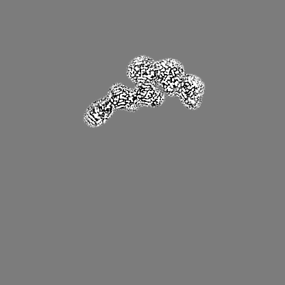
| Title | Focus refinement map of PSII-CAC | |||||||||
 Map data Map data | Focus refinement map of 6CAC of PSII-CAC from Rhodomonas salina | |||||||||
 Sample Sample |
| |||||||||
 Keywords Keywords | Photosystem II associated with CAC antenna from Rhodomonas Salina / PHOTOSYNTHESIS | |||||||||
| Biological species |  Rhodomonas salina (eukaryote) Rhodomonas salina (eukaryote) | |||||||||
| Method | single particle reconstruction / cryo EM / Resolution: 2.9 Å | |||||||||
 Authors Authors | Si L / Li M | |||||||||
| Funding support |  China, 1 items China, 1 items
| |||||||||
 Citation Citation | Journal: Nat Commun / Year: 2024 Title: Structural basis for the distinct core-antenna assembly of cryptophyte photosystem II. Authors: Long Si / Shumeng Zhang / Xiaodong Su / Mei Li / Abstract: Photosystem II (PSII) catalyzes the light-driven charge separation and water oxidation reactions of photosynthesis. Eukaryotic PSII core is usually associated with membrane-embedded light-harvesting ...Photosystem II (PSII) catalyzes the light-driven charge separation and water oxidation reactions of photosynthesis. Eukaryotic PSII core is usually associated with membrane-embedded light-harvesting antennae, which greatly increase the absorbance cross-section of the core. The peripheral antennae in different phototrophs vary considerably in protein composition and arrangement. Photosynthetic cryptophytes possess chlorophyll a/c binding proteins (CACs) that serve as their antennae. How these CACs assemble with the PSII core remains unclear. Here, we report the 2.57-Å resolution structure of cryptophyte PSII-CAC purified from cells at nitrogen-limited stationary growth phase. We show that each monomer of the PSII homodimer contains a core complex, six chlorophyll a/c binding proteins (CACs) and a previously unseen chlorophyll-binding protein (termed CAL-II). Six CACs are arranged as a double-layered arc-shaped non-parallel belt, and two such belts attach to the dimeric core from opposite sides. The CAL-II simultaneously interacts with a number of core subunits and five CACs. The distinct organization of CACs and the presence of CAL-II may play a critical role in stabilizing the dimeric PSII-CAC complex under stress conditions. Our study provides mechanistic insights into the assembly and function of the PSII-CAC complex as well as the possible adaptation of cryptophytes in response to environmental stresses. | |||||||||
| History |
|
- Structure visualization
| Supplemental images |
|---|
- Downloads & links
-EMDB archive
| Map data | emd_38913.map.gz | 15.5 MB |  EMDB map data format EMDB map data format | |
|---|---|---|---|---|
| Header (meta data) | emd-38913-v30.xmlemd-38913.xml | 12.2 KB 12.2 KB | Display Display | EMDB header |
| Images |  emd_38913.png emd_38913.png | 35.7 KB | ||
| Filedesc metadata | emd-38913.cif.gz | 4 KB | ||
| Others | emd_38913_half_map_1.map.gzemd_38913_half_map_2.map.gz | 218.5 MB 218.7 MB | ||
| Archive directory |  http://ftp.pdbj.org/pub/emdb/structures/EMD-38913ftp://ftp.pdbj.org/pub/emdb/structures/EMD-38913 http://ftp.pdbj.org/pub/emdb/structures/EMD-38913ftp://ftp.pdbj.org/pub/emdb/structures/EMD-38913 | HTTPS FTP |
-Related structure data
-Links
| EMDB pages | EMDB (EBI/PDBe) / EMDataResource |
|---|
-Map
| File | Download / File: emd_38913.map.gz / Format: CCP4 / Size: 274.6 MB / Type: IMAGE STORED AS FLOATING POINT NUMBER (4 BYTES) | ||||||||||||||||||||||||||||||||||||
|---|---|---|---|---|---|---|---|---|---|---|---|---|---|---|---|---|---|---|---|---|---|---|---|---|---|---|---|---|---|---|---|---|---|---|---|---|---|
| Annotation | Focus refinement map of 6CAC of PSII-CAC from Rhodomonas salina | ||||||||||||||||||||||||||||||||||||
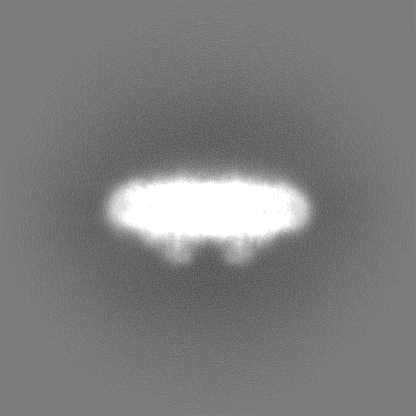
| Projections & slices | Image control
Images are generated by Spider. | ||||||||||||||||||||||||||||||||||||
| Voxel size | X=Y=Z: 1.04003 Å | ||||||||||||||||||||||||||||||||||||
| Density |
| ||||||||||||||||||||||||||||||||||||
| Symmetry | Space group: 1 | ||||||||||||||||||||||||||||||||||||
| Details | EMDB XML:
|
 Z (Sec.)
 Y (Row.)
 X (Col.)


-Supplemental data
-Half map: #1
| File | emd_38913_half_map_1.map | ||||||||||||
|---|---|---|---|---|---|---|---|---|---|---|---|---|---|
| Projections & Slices |
| ||||||||||||
| Density Histograms |


-Half map: #2
| File | emd_38913_half_map_2.map | ||||||||||||
|---|---|---|---|---|---|---|---|---|---|---|---|---|---|
| Projections & Slices |
| ||||||||||||
| Density Histograms |


- Sample components
-Entire : Inactive Photosystem II associated with chlorophyll a/c binding p...
| Entire | Name: Inactive Photosystem II associated with chlorophyll a/c binding protein |
|---|---|
| Components |
|
-Supramolecule #1: Inactive Photosystem II associated with chlorophyll a/c binding p...
| Supramolecule | Name: Inactive Photosystem II associated with chlorophyll a/c binding protein type: complex / ID: 1 / Parent: 0 / Macromolecule list: all |
|---|---|
| Source (natural) | Organism: Rhodomonas salina (eukaryote) |
-Macromolecule #1: CAC antenna
| Macromolecule | Name: CAC antenna / type: other / ID: 1 / Classification: other |
|---|---|
| Source (natural) | Organism: Rhodomonas salina (eukaryote) |
| Sequence | String: NO |
-Experimental details
-Structure determination
| Method | cryo EM |
|---|---|
 Processing Processing | single particle reconstruction |
| Aggregation state | 3D array |
-Sample preparation
| Buffer | pH: 7.5 |
|---|---|
| Vitrification | Cryogen name: ETHANE |
- Electron microscopy
| Microscope | FEI TITAN |
|---|---|
| Image recording | Film or detector model: DIRECT ELECTRON DE-16 (4k x 4k) / Average electron dose: 60.0 e/Å2 |
| Electron beam | Acceleration voltage: 300 kV / Electron source:  FIELD EMISSION GUN FIELD EMISSION GUN |
| Electron optics | Illumination mode: FLOOD BEAM / Imaging mode: BRIGHT FIELD / Nominal defocus max: 2.2 µm / Nominal defocus min: 1.2 µm |
-Image processing
| Startup model | Type of model: PDB ENTRY PDB model - PDB ID: |
|---|---|
| Final reconstruction | Resolution.type: BY AUTHOR / Resolution: 2.9 Å / Resolution method: FSC 0.143 CUT-OFF / Number images used: 123227 |
| Initial angle assignment | Type: ANGULAR RECONSTITUTION |
| Final angle assignment | Type: ANGULAR RECONSTITUTION |

