National Natural Science Foundation of China (NSFC)
China
Citation
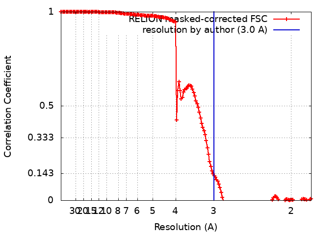
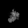
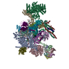
Journal: EMBO J / Year: 2025 Title: Structure of a step II catalytically activated spliceosome from Chlamydomonas reinhardtii. Authors: Yichen Lu / Ke Liang / Xiechao Zhan / Abstract: Pre-mRNA splicing, a fundamental step in eukaryotic gene expression, is executed by the spliceosomes. While there is extensive knowledge of the composition and structure of spliceosomes in yeasts and ...Pre-mRNA splicing, a fundamental step in eukaryotic gene expression, is executed by the spliceosomes. While there is extensive knowledge of the composition and structure of spliceosomes in yeasts and humans, the structural diversity of spliceosomes in non-canonical organisms remains unclear. Here, we present a cryo-EM structure of a step II catalytically activated spliceosome (C complex) derived from the unicellular green alga Chlamydomonas reinhardtii at 2.6 Å resolution. This Chlamydomonas C complex comprises 29 proteins and four RNA elements, creating a dynamic assembly that shares a similar overall architecture with yeast and human counterparts but also has unique features of its own. Distinctive structural characteristics include variations in protein compositions as well as some noteworthy RNA features. The splicing factor Prp17, with four fragments and a WD40 domain, is engaged in intricate interactions with multiple protein and RNA components. The structural elucidation of Chlamydomonas C complex provides insights into the molecular mechanism of RNA splicing in plants and understanding splicing evolution in eukaryotes.
In the structure databanks used in Yorodumi, some data are registered as the other names, "COVID-19 virus" and "2019-nCoV". Here are the details of the virus and the list of structure data.
Jan 31, 2019. EMDB accession codes are about to change! (news from PDBe EMDB page)
EMDB accession codes are about to change! (news from PDBe EMDB page)
The allocation of 4 digits for EMDB accession codes will soon come to an end. Whilst these codes will remain in use, new EMDB accession codes will include an additional digit and will expand incrementally as the available range of codes is exhausted. The current 4-digit format prefixed with “EMD-” (i.e. EMD-XXXX) will advance to a 5-digit format (i.e. EMD-XXXXX), and so on. It is currently estimated that the 4-digit codes will be depleted around Spring 2019, at which point the 5-digit format will come into force.
The EM Navigator/Yorodumi systems omit the EMD- prefix.
Related info.:Q: What is EMD? / ID/Accession-code notation in Yorodumi/EM Navigator
Yorodumi is a browser for structure data from EMDB, PDB, SASBDB, etc.
This page is also the successor to EM Navigator detail page, and also detail information page/front-end page for Omokage search.
The word "yorodu" (or yorozu) is an old Japanese word meaning "ten thousand". "mi" (miru) is to see.
Related info.:EMDB / PDB / SASBDB / Comparison of 3 databanks / Yorodumi Search / Aug 31, 2016. New EM Navigator & Yorodumi / Yorodumi Papers / Jmol/JSmol / Function and homology information / Changes in new EM Navigator and Yorodumi
 Movie
Movie Controller

 Yorodumi
 Open data
 Basic information
 Map data
 Sample
 Keywords
 Chlamydomonas reinhardtii (plant)
 Authors
 China, 1 items
  Citation
 Structure visualization
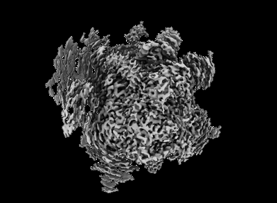
 Downloads & links
 EMDB map data format
 emd_38364.png
 http://ftp.pdbj.org/pub/emdb/structures/EMD-38364
 Z (Sec.)
 Y (Row.)
 X (Col.)


 Sample components
 Processing
 Electron microscopy
 FIELD EMISSION GUN