Journal: ACS Pharmacol Transl Sci / Year: 2025 Title: Aglycone Polyether Ionophores Affecting Actin Filaments as Broad-Spectrum Antiviral Agents. Authors: Minjian Huang / Jiaqi Li / Jing Li / Ben Hu / Ran Liu / Lianghao Huang / Caiqian Wang / Rong Hua / Chuanjian Wu / Zhuli Li / Zherui Zhang / Yanan Zhang / Yingjun Wu / Qiuyan Zhang / Yong ...Authors: Minjian Huang / Jiaqi Li / Jing Li / Ben Hu / Ran Liu / Lianghao Huang / Caiqian Wang / Rong Hua / Chuanjian Wu / Zhuli Li / Zherui Zhang / Yanan Zhang / Yingjun Wu / Qiuyan Zhang / Yong Wang / Jing Liu / Zixin Deng / Wei Wang / Wei Hou / Ling Zhao / Yucheng Xia / Xiaolian Zhang / Longfei Wang / Bo Zhang / Tiangang Liu / Abstract: RNA viruses have high mutation rates and constitute an increasing global risk. As the viral target approach to develop antiviral drugs is inadequate for responding to an increasing diversity of ...RNA viruses have high mutation rates and constitute an increasing global risk. As the viral target approach to develop antiviral drugs is inadequate for responding to an increasing diversity of viruses, an urgent need exists for the development of new antivirals to prevent future outbreaks. Here, we show that aglycone ionophores maduramycin (Mad) and endusamycin (End) from are broadly virucidal against cytoplasmic replicated viruses, including Japanese encephalitis virus (JEV), rabies virus, hepatitis C virus, vesicular stomatitis virus, hantavirus, dengue virus, Zika virus, chikungunya virus, and SARS-CoV-2 in vitro. Mechanistic studies suggest Mad and End can target actin filaments and displace the DNase-I-binding loop (D-loop) into an outward conformation for stabilizing actin filaments and primarily inhibit viral replication. Liposome-encapsulated Mad or End fully protects mice against JEV infection in vivo. Thus, our results may provide potential and naturally produced antivirals to prevent the spread of viruses in animals.
In the structure databanks used in Yorodumi, some data are registered as the other names, "COVID-19 virus" and "2019-nCoV". Here are the details of the virus and the list of structure data.
Jan 31, 2019. EMDB accession codes are about to change! (news from PDBe EMDB page)
EMDB accession codes are about to change! (news from PDBe EMDB page)
The allocation of 4 digits for EMDB accession codes will soon come to an end. Whilst these codes will remain in use, new EMDB accession codes will include an additional digit and will expand incrementally as the available range of codes is exhausted. The current 4-digit format prefixed with “EMD-” (i.e. EMD-XXXX) will advance to a 5-digit format (i.e. EMD-XXXXX), and so on. It is currently estimated that the 4-digit codes will be depleted around Spring 2019, at which point the 5-digit format will come into force.
The EM Navigator/Yorodumi systems omit the EMD- prefix.
Related info.:Q: What is EMD? / ID/Accession-code notation in Yorodumi/EM Navigator
Yorodumi is a browser for structure data from EMDB, PDB, SASBDB, etc.
This page is also the successor to EM Navigator detail page, and also detail information page/front-end page for Omokage search.
The word "yorodu" (or yorozu) is an old Japanese word meaning "ten thousand". "mi" (miru) is to see.
Related info.:EMDB / PDB / SASBDB / Comparison of 3 databanks / Yorodumi Search / Aug 31, 2016. New EM Navigator & Yorodumi / Yorodumi Papers / Jmol/JSmol / Function and homology information / Changes in new EM Navigator and Yorodumi
 Movie
Movie Controller


 Open data
 Basic information
 Map data
 Sample
 Keywords
 Function and homology information
 Authors
 Citation
 Structure visualization
 Downloads & links
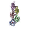
 emd_38281.png
 http://ftp.pdbj.org/pub/emdb/structures/EMD-38281


 Z (Sec.)
 Y (Row.)
 X (Col.)


 Sample components
 Processing
 Electron microscopy
 FIELD EMISSION GUN