Movie
Movie Controller

[English] 日本語
 Yorodumi
- EMDB-38080: SIRM reconstruction of the MC-45 de novo processed ribosome 50S -
+ Open data

- Basic information
| Entry | 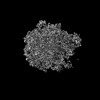 | |||||||||
|---|---|---|---|---|---|---|---|---|---|---|
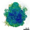
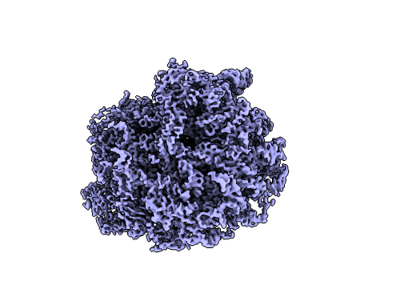
| Title | SIRM reconstruction of the MC-45 de novo processed ribosome 50S | |||||||||
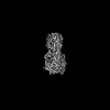
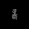
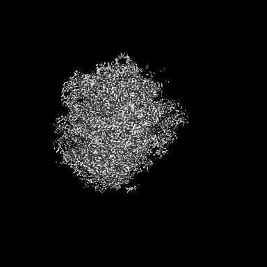
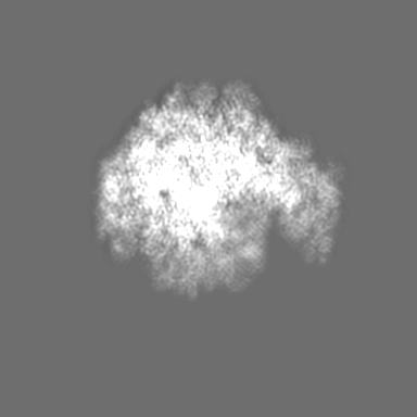
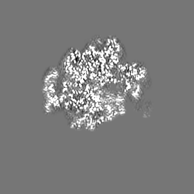
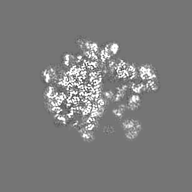
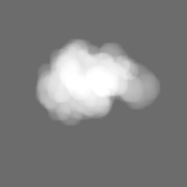
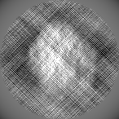
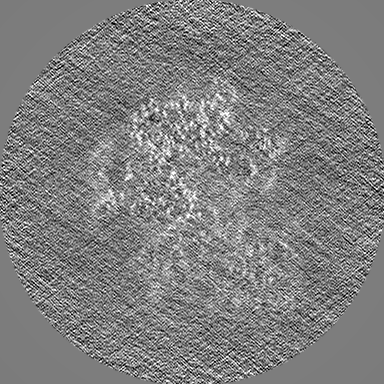
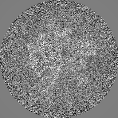
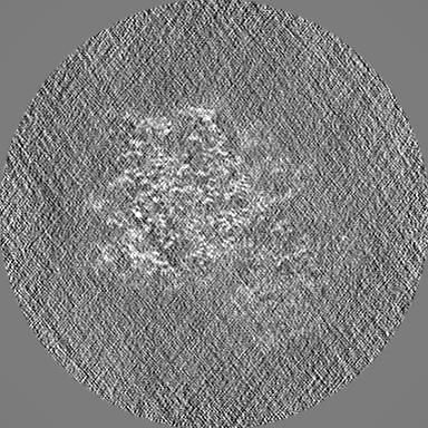
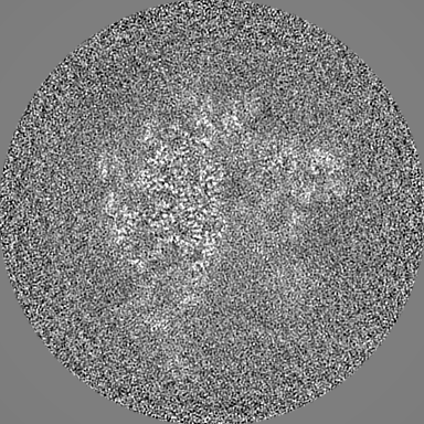
 Map data Map data | The SIRM reconstructed ribosome 50S. The map was low-passed to 3.6 Angstrom. This map was reconstructed after Validation and Mask-Picking. | |||||||||
 Sample Sample |
| |||||||||
 Keywords Keywords | ribosome 50S / RIBOSOME | |||||||||
| Biological species |  | |||||||||
| Method | single particle reconstruction / Resolution: 3.6 Å | |||||||||
 Authors Authors | Zhang XZ / Zhu DJ / Cao WL | |||||||||
| Funding support |  China, 1 items China, 1 items
| |||||||||
 Citation Citation | Journal: Sci Adv / Year: 2024 Title: Correction of preferred orientation-induced distortion in cryo-electron microscopy maps. Authors: Dongjie Zhu / Weili Cao / Junxi Li / Chunling Wu / Duanfang Cao / Xinzheng Zhang / Abstract: Reconstruction maps of cryo-electron microscopy (cryo-EM) exhibit distortion when the cryo-EM dataset is incomplete, usually caused by unevenly distributed orientations. Prior efforts had been ...Reconstruction maps of cryo-electron microscopy (cryo-EM) exhibit distortion when the cryo-EM dataset is incomplete, usually caused by unevenly distributed orientations. Prior efforts had been attempted to address this preferred orientation problem using tilt-collection strategy and modifications to grids or to air-water interfaces. However, these approaches often require time-consuming experiments, and the effect was always protein dependent. Here, we developed a procedure containing removing misaligned particles and an iterative reconstruction method based on signal-to-noise ratio of Fourier component to correct this distortion by recovering missing data using a purely computational algorithm. This procedure called signal-to-noise ratio iterative reconstruction method (SIRM) was applied on incomplete datasets of various proteins to fix distortion in cryo-EM maps and to a more isotropic resolution. In addition, SIRM provides a better reference map for further reconstruction refinements, resulting in an improved alignment, which ultimately improves map quality and benefits model building. | |||||||||
| History |
|
- Structure visualization
| Supplemental images |
|---|
- Downloads & links
-EMDB archive
| Map data | emd_38080.map.gz | 20.3 MB |  EMDB map data format EMDB map data format | |
|---|---|---|---|---|
| Header (meta data) | emd-38080-v30.xmlemd-38080.xml | 13 KB 13 KB | Display Display | EMDB header |
| Images |  emd_38080.png emd_38080.png | 78.9 KB | ||
| Masks | emd_38080_msk_1.map | 216 MB | Mask map | |
| Filedesc metadata | emd-38080.cif.gz | 3.8 KB | ||
| Others | emd_38080_half_map_1.map.gzemd_38080_half_map_2.map.gz | 172.3 MB 172.2 MB | ||
| Archive directory |  http://ftp.pdbj.org/pub/emdb/structures/EMD-38080ftp://ftp.pdbj.org/pub/emdb/structures/EMD-38080 http://ftp.pdbj.org/pub/emdb/structures/EMD-38080ftp://ftp.pdbj.org/pub/emdb/structures/EMD-38080 | HTTPS FTP |
-Related structure data


-Links
| EMDB pages | EMDB (EBI/PDBe) / EMDataResource |
|---|
-Map
| File | Download / File: emd_38080.map.gz / Format: CCP4 / Size: 216 MB / Type: IMAGE STORED AS FLOATING POINT NUMBER (4 BYTES) | ||||||||||||||||||||||||||||||||||||
|---|---|---|---|---|---|---|---|---|---|---|---|---|---|---|---|---|---|---|---|---|---|---|---|---|---|---|---|---|---|---|---|---|---|---|---|---|---|
| Annotation | The SIRM reconstructed ribosome 50S. The map was low-passed to 3.6 Angstrom. This map was reconstructed after Validation and Mask-Picking. | ||||||||||||||||||||||||||||||||||||
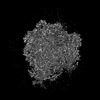
| Projections & slices | Image control
Images are generated by Spider. | ||||||||||||||||||||||||||||||||||||
| Voxel size | X=Y=Z: 0.94666 Å | ||||||||||||||||||||||||||||||||||||
| Density |
| ||||||||||||||||||||||||||||||||||||
| Symmetry | Space group: 1 | ||||||||||||||||||||||||||||||||||||
| Details | EMDB XML:
|
 Z (Sec.)
 Y (Row.)
 X (Col.)


-Supplemental data
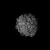
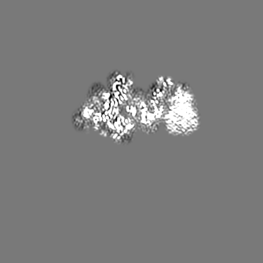
-Mask #1
| File | emd_38080_msk_1.map | ||||||||||||
|---|---|---|---|---|---|---|---|---|---|---|---|---|---|
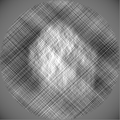
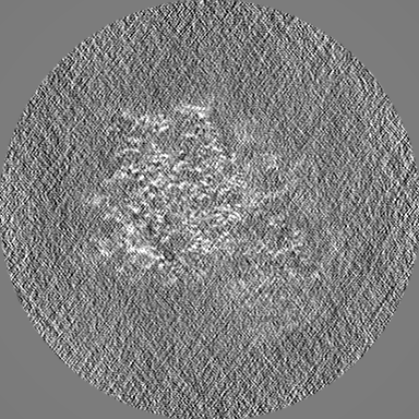
| Projections & Slices |
| ||||||||||||
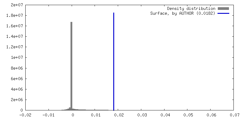
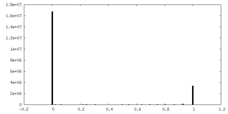
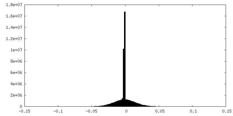
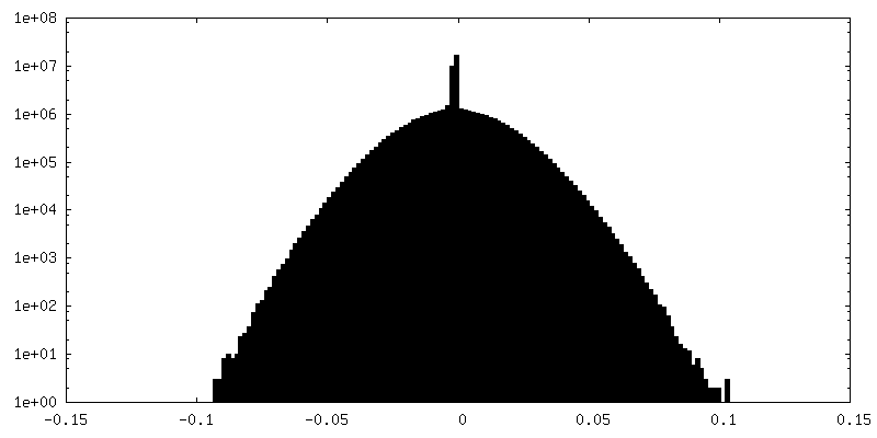
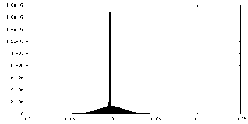
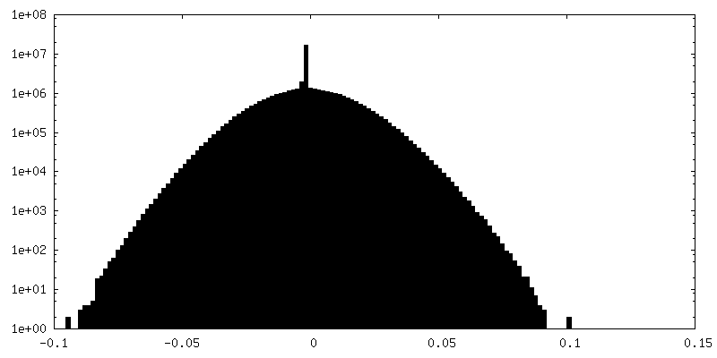
| Density Histograms |


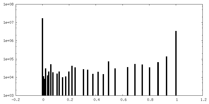
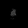
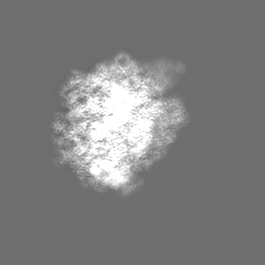
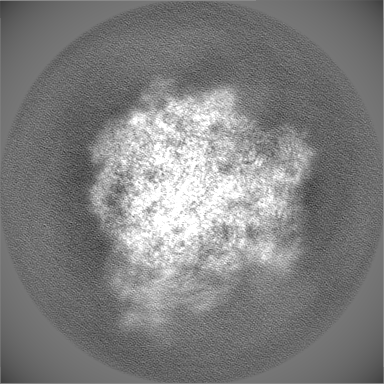
-Half map: These two unfil-maps were NOT reconstructed by SIRM....
| File | emd_38080_half_map_1.map | ||||||||||||
|---|---|---|---|---|---|---|---|---|---|---|---|---|---|
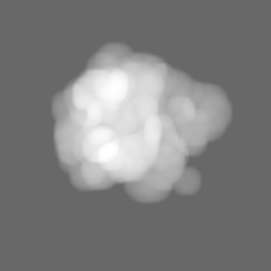
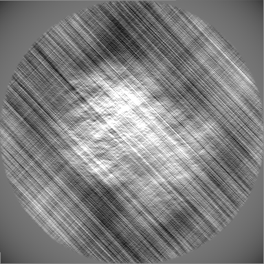
| Annotation | These two unfil-maps were NOT reconstructed by SIRM. They were reconstructed by conventional relion_reconstruct, to generate a FSC curve. | ||||||||||||
| Projections & Slices |
| ||||||||||||
| Density Histograms |
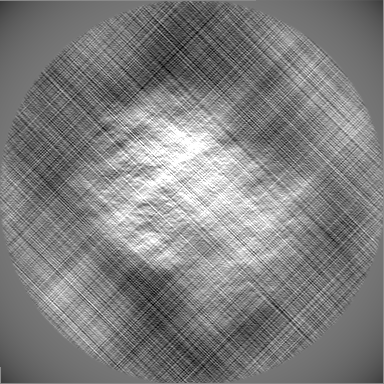
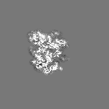
-Half map: These two unfil-maps were NOT reconstructed by SIRM....
| File | emd_38080_half_map_2.map | ||||||||||||
|---|---|---|---|---|---|---|---|---|---|---|---|---|---|
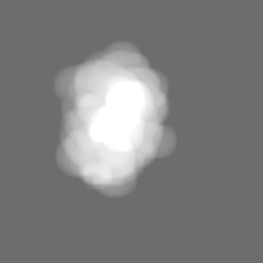
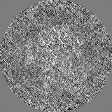
| Annotation | These two unfil-maps were NOT reconstructed by SIRM. They were reconstructed by conventional relion_reconstruct, to generate a FSC curve. | ||||||||||||
| Projections & Slices |
| ||||||||||||
| Density Histograms |

- Sample components
-Entire : E. coli 70S ribosome, from EMPIAR-10509
| Entire | Name: E. coli 70S ribosome, from EMPIAR-10509 |
|---|---|
| Components |
|
-Supramolecule #1: E. coli 70S ribosome, from EMPIAR-10509
| Supramolecule | Name: E. coli 70S ribosome, from EMPIAR-10509 / type: complex / ID: 1 / Parent: 0 |
|---|---|
| Source (natural) | Organism: |
-Experimental details
-Structure determination
 Processing Processing | single particle reconstruction |
|---|---|
| Aggregation state | particle |
-Sample preparation
| Buffer | pH: 7.5 |
|---|
- Electron microscopy
| Microscope | FEI TITAN KRIOS |
|---|---|
| Image recording | Film or detector model: GATAN K3 (6k x 4k) / Average electron dose: 40.0 e/Å2 Details: We used EMPIAR-10509. This repository was not recorded by us. |
| Electron beam | Acceleration voltage: 300 kV / Electron source:  FIELD EMISSION GUN FIELD EMISSION GUN |
| Electron optics | Illumination mode: FLOOD BEAM / Imaging mode: BRIGHT FIELD / Nominal defocus max: 1.5 µm / Nominal defocus min: 0.8 µm |
| Experimental equipment |  Model: Titan Krios / Image courtesy: FEI Company |
-Image processing
| Startup model | Type of model: EMDB MAP EMDB ID: |
|---|---|
| Final reconstruction | Applied symmetry - Point group: C1 (asymmetric) / Resolution.type: BY AUTHOR / Resolution: 3.6 Å / Resolution method: OTHER / Software - Name: RELION (ver. 3.1.2) / Software - details: SIRM-RELION Details: The map was low-passed to 3.6 Angstrom. The reported resolution was 3.1 Angstrom via FSC-0.143 (Gold-standard). Number images used: 28050 |
| Initial angle assignment | Type: MAXIMUM LIKELIHOOD |
| Final angle assignment | Type: MAXIMUM LIKELIHOOD / Software - Name: RELION (ver. 3.1.2) |