Movie
Movie Controller


+ Open data

- Basic information
| Entry |  | |||||||||
|---|---|---|---|---|---|---|---|---|---|---|
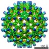
| Title | Hepatitis B virus capsid in complex with SR protein kinase 2 | |||||||||
 Map data Map data | ||||||||||
 Sample Sample |
| |||||||||
 Keywords Keywords | Viral core capsid / Icosahedral / Kinase / Complex / VIRUS | |||||||||
| Biological species |  Homo sapiens (human) / Homo sapiens (human) /   Hepatitis B virus Hepatitis B virus | |||||||||
| Method | single particle reconstruction / cryo EM / Resolution: 4.6 Å | |||||||||
 Authors Authors | Yip RPH / Lai LTF / Kwok DCY / Lau WCY / Ngo JCK | |||||||||
| Funding support |  Hong Kong, 1 items Hong Kong, 1 items
| |||||||||
 Citation Citation | Journal: PLoS Pathog / Year: 2024 Title: SRPK2 Mediates HBV Core Protein Phosphorylation and Capsid Assembly via Docking Interaction. Authors: Ryan Pak Hong Yip / Doris Ching Ying Kwok / Louis Tung Faat Lai / Siu-Ming Ho / Ivan Chun Kit Wong / Chi-Ping Chan / Wilson Chun Yu Lau / Jacky Chi Ki Ngo / Abstract: Members of the serine-arginine protein kinase (SRPK) family, SRPK1 and SRPK2, phosphorylate the hepatitis B core protein (Cp) and are crucial for pregenomic RNA encapsidation during viral ...Members of the serine-arginine protein kinase (SRPK) family, SRPK1 and SRPK2, phosphorylate the hepatitis B core protein (Cp) and are crucial for pregenomic RNA encapsidation during viral nucleocapsid assembly. Among them, SRPK2 exhibits higher kinase activity toward Cp. In this study, we identified Cp sites that are phosphorylated by SRPK2 and demonstrated that the kinase utilizes an SRPK-specific docking groove to interact with and regulate the phosphorylation of the C-terminal arginine rich domain of Cp. We determined that direct interaction between the docking groove of SRPK2 and unphosphorylated Cp inhibited premature viral capsid assembly in vitro, whereas the phosphorylation of the viral protein reactivated the process. Pull-down assays together with the new cryo-electron microscopy structure of the HBV capsid in complex with SRPK2 revealed that the kinases decorate the surface of the viral capsid by interacting with the C-terminal domain of Cp, underscoring the importance of the docking interaction in regulating capsid assembly and pregenome packaging. Moreover, SRPK2-knockout in HepG2 cells suppressed Cp phosphorylation, indicating that SRPK2 is an important cellular kinase for HBV life cycle. | |||||||||
| History |
|
- Structure visualization
| Supplemental images |
|---|

- Downloads & links
-EMDB archive
| Map data | emd_38062.map.gz | 35.9 MB |  EMDB map data format EMDB map data format | |
|---|---|---|---|---|
| Header (meta data) | emd-38062-v30.xmlemd-38062.xml | 17.5 KB 17.5 KB | Display Display | EMDB header |
| FSC (resolution estimation) | emd_38062_fsc.xml | 9.1 KB | Display | FSC data file |
| Images | 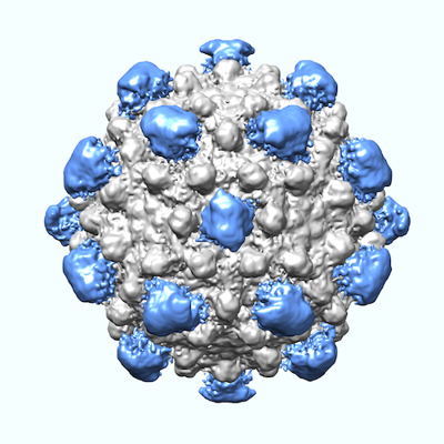 emd_38062.png emd_38062.png | 154 KB | ||
| Masks | emd_38062_msk_1.map | 64 MB | Mask map | |
| Filedesc metadata | emd-38062.cif.gz | 5.3 KB | ||
| Others | emd_38062_half_map_1.map.gzemd_38062_half_map_2.map.gz | 49.3 MB 49.4 MB | ||
| Archive directory |  http://ftp.pdbj.org/pub/emdb/structures/EMD-38062ftp://ftp.pdbj.org/pub/emdb/structures/EMD-38062 http://ftp.pdbj.org/pub/emdb/structures/EMD-38062ftp://ftp.pdbj.org/pub/emdb/structures/EMD-38062 | HTTPS FTP |
-Related structure data

-Links
| EMDB pages | EMDB (EBI/PDBe) / EMDataResource |
|---|
-Map
| File | Download / File: emd_38062.map.gz / Format: CCP4 / Size: 64 MB / Type: IMAGE STORED AS FLOATING POINT NUMBER (4 BYTES) | ||||||||||||||||||||||||||||||||||||
|---|---|---|---|---|---|---|---|---|---|---|---|---|---|---|---|---|---|---|---|---|---|---|---|---|---|---|---|---|---|---|---|---|---|---|---|---|---|
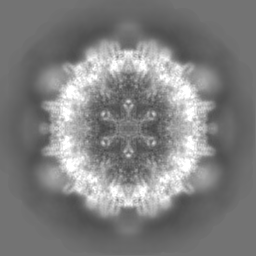
| Projections & slices | Image control
Images are generated by Spider. | ||||||||||||||||||||||||||||||||||||
| Voxel size | X=Y=Z: 1.938 Å | ||||||||||||||||||||||||||||||||||||
| Density |
| ||||||||||||||||||||||||||||||||||||
| Symmetry | Space group: 1 | ||||||||||||||||||||||||||||||||||||
| Details | EMDB XML:
|
 Z (Sec.)
 Y (Row.)
 X (Col.)


-Supplemental data
-Mask #1
| File | emd_38062_msk_1.map | ||||||||||||
|---|---|---|---|---|---|---|---|---|---|---|---|---|---|
| Projections & Slices |
| ||||||||||||
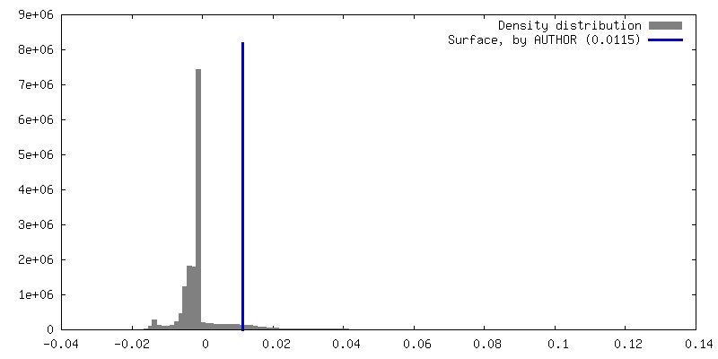
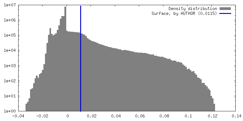
| Density Histograms |


-Half map: #1
| File | emd_38062_half_map_1.map | ||||||||||||
|---|---|---|---|---|---|---|---|---|---|---|---|---|---|
| Projections & Slices |
| ||||||||||||
| Density Histograms |


-Half map: #2
| File | emd_38062_half_map_2.map | ||||||||||||
|---|---|---|---|---|---|---|---|---|---|---|---|---|---|
| Projections & Slices |
| ||||||||||||
| Density Histograms |


- Sample components
-Entire : Hepatitis B virus capsid in complex with SR protein kinase 2
| Entire | Name: Hepatitis B virus capsid in complex with SR protein kinase 2 |
|---|---|
| Components |
|
-Supramolecule #1: Hepatitis B virus capsid in complex with SR protein kinase 2
| Supramolecule | Name: Hepatitis B virus capsid in complex with SR protein kinase 2 type: complex / ID: 1 / Parent: 0 / Macromolecule list: all |
|---|---|
| Source (natural) | Organism: Homo sapiens (human) |
| Molecular weight | Theoretical: 52 kDa/nm |
-Supramolecule #2: SR protein kinase 2
| Supramolecule | Name: SR protein kinase 2 / type: complex / ID: 2 / Parent: 1 / Macromolecule list: #1 |
|---|---|
| Source (natural) | Organism: Hepatitis B virus |
-Supramolecule #3: Hepatitis B virus capsid
| Supramolecule | Name: Hepatitis B virus capsid / type: complex / ID: 3 / Parent: 1 / Macromolecule list: #2 |
|---|
-Macromolecule #1: SR protein kinase 2
| Macromolecule | Name: SR protein kinase 2 / type: protein_or_peptide / ID: 1 / Enantiomer: LEVO |
|---|---|
| Source (natural) | Organism: Homo sapiens (human) |
| Recombinant expression | Organism:  |
| Sequence | String: MGSSHHHHHH SSGLVPRGSH NMSVNSEKSS SSERPEPQQK APLVPPPPPP PPPPPPPLPD PTPPEPEEEI LGSDDEEQED PADYCKGGYH PVKIGDLFNG RYHVIRKLGW GHFSTVWLCW DMQGKRFVAM KVVKSAQHYT ETALDEIKLL KCVRESDPSD PNKDMVVQLI ...String: MGSSHHHHHH SSGLVPRGSH NMSVNSEKSS SSERPEPQQK APLVPPPPPP PPPPPPPLPD PTPPEPEEEI LGSDDEEQED PADYCKGGYH PVKIGDLFNG RYHVIRKLGW GHFSTVWLCW DMQGKRFVAM KVVKSAQHYT ETALDEIKLL KCVRESDPSD PNKDMVVQLI DDFKISGMNG IHVCMVFEVL GHHLLKWIIK SNYQGLPVRC VKSIIRQVLQ GLDYLHSKCK IIHTDIKPEN ILMCVDDAYV RRMAAEATEW QKAGAPPPSG SAVSTAPRAA DLLVNPLDPR NADKIRVKIA DLGNACWVHK HFTEDIQTRQ YRSIEVLIGA GYSTPADIWS TACMAFELAT GDYLFEPHSG EDYSRDEDHI AHIIELLGSI PRHFALSGKY SREFFNRRGE LRHITKLKPW SLFDVLVEKY GWPHEDAAQF TDFLIPMLEM VPEKRASAGE CLRHPWLNS |
-Macromolecule #2: Hepatitis B virus core protein
| Macromolecule | Name: Hepatitis B virus core protein / type: protein_or_peptide / ID: 2 / Enantiomer: LEVO |
|---|---|
| Source (natural) | Organism: Hepatitis B virus |
| Recombinant expression | Organism: |
| Sequence | String: MGSSHHHHHH SSGLVPRGSH MDIDPYKEFG ATVELLSFLP SDFFPSVRDL LDTASALYRE ALESPEHCSP HHTALRQAIL CWGELMTLAT WVGNNLEDPA SRDLVVNYVN TNMGLKIRQL LWFHISCLTF GRETVLEYLV SFGVWIRTPP AYRPPNAPIL STLPETTVVR ...String: MGSSHHHHHH SSGLVPRGSH MDIDPYKEFG ATVELLSFLP SDFFPSVRDL LDTASALYRE ALESPEHCSP HHTALRQAIL CWGELMTLAT WVGNNLEDPA SRDLVVNYVN TNMGLKIRQL LWFHISCLTF GRETVLEYLV SFGVWIRTPP AYRPPNAPIL STLPETTVVR RRDRGRSPRR RTPSPRRRRS QSPRRRRSQS RESQC |
-Experimental details
-Structure determination
| Method | cryo EM |
|---|---|
 Processing Processing | single particle reconstruction |
| Aggregation state | particle |
-Sample preparation
| Concentration | 0.9 mg/mL |
|---|---|
| Buffer | pH: 7.4 |
| Vitrification | Cryogen name: ETHANE / Chamber humidity: 100 % / Chamber temperature: 277.15 K / Instrument: FEI VITROBOT MARK IV |
- Electron microscopy
| Microscope | TFS TALOS F200C |
|---|---|
| Image recording | Film or detector model: FEI FALCON III (4k x 4k) / Detector mode: COUNTING / Number real images: 824 / Average exposure time: 54.0 sec. / Average electron dose: 50.0 e/Å2 |
| Electron beam | Acceleration voltage: 200 kV / Electron source:  FIELD EMISSION GUN FIELD EMISSION GUN |
| Electron optics | Illumination mode: FLOOD BEAM / Imaging mode: BRIGHT FIELD / Cs: 2.7 mm / Nominal defocus max: 3.0 µm / Nominal defocus min: 0.5 µm / Nominal magnification: 150000 |
| Sample stage | Specimen holder model: GATAN 626 SINGLE TILT LIQUID NITROGEN CRYO TRANSFER HOLDER Cooling holder cryogen: NITROGEN |
| Experimental equipment |  Model: Tecnai F20 / Image courtesy: FEI Company |