 Movie
Movie Controller

[English] 日本語
 Yorodumi
- EMDB-3793: Cryotomogram of hexagonal arrays of contractile injection systems -
+ Open data

- Basic information
| Entry | Database: EMDB / ID: EMD-3793 | |||||||||
|---|---|---|---|---|---|---|---|---|---|---|
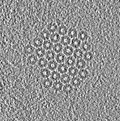
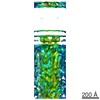
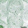
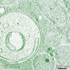
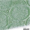
| Title | Cryotomogram of hexagonal arrays of contractile injection systems | |||||||||
 Map data Map data | Hexagonal arrays of contractile injection systems | |||||||||
 Sample Sample |
| |||||||||
| Biological species |  Candidatus Amoebophilus asiaticus 5a2 (bacteria) Candidatus Amoebophilus asiaticus 5a2 (bacteria) | |||||||||
| Method | electron tomography / cryo EM | |||||||||
 Authors Authors | Boeck D / Medeiros JM | |||||||||
 Citation Citation | Journal: Science / Year: 2017 Title: In situ architecture, function, and evolution of a contractile injection system. Authors: Désirée Böck / João M Medeiros / Han-Fei Tsao / Thomas Penz / Gregor L Weiss / Karin Aistleitner / Matthias Horn / Martin Pilhofer /   Abstract: Contractile injection systems mediate bacterial cell-cell interactions by a bacteriophage tail-like structure. In contrast to extracellular systems, the type 6 secretion system (T6SS) is defined by ...Contractile injection systems mediate bacterial cell-cell interactions by a bacteriophage tail-like structure. In contrast to extracellular systems, the type 6 secretion system (T6SS) is defined by intracellular localization and attachment to the cytoplasmic membrane. Here we used cryo-focused ion beam milling, electron cryotomography, and functional assays to study a T6SS in The in situ architecture revealed three modules, including a contractile sheath-tube, a baseplate, and an anchor. All modules showed conformational changes upon firing. Lateral baseplate interactions coordinated T6SSs in hexagonal arrays. The system mediated interactions with host membranes and may participate in phagosome escape. Evolutionary sequence analyses predicted that T6SSs are more widespread than previously thought. Our insights form the basis for understanding T6SS key concepts and exploring T6SS diversity. | |||||||||
| History |
|
- Structure visualization
| Movie |
 Movie viewer Movie viewer |
|---|---|
| Structure viewer | EM map: SurfViewMolmilJmol/JSmol |
| Supplemental images |
- Downloads & links
-EMDB archive
| Map data | emd_3793.map.gz | 220 MB | EMDB map data format | |
|---|---|---|---|---|
| Header (meta data) | emd-3793-v30.xmlemd-3793.xml | 7.1 KB 7.1 KB | Display Display | EMDB header |
| Images |  emd_3793.png emd_3793.png | 141 KB | ||
| Archive directory |  http://ftp.pdbj.org/pub/emdb/structures/EMD-3793ftp://ftp.pdbj.org/pub/emdb/structures/EMD-3793 http://ftp.pdbj.org/pub/emdb/structures/EMD-3793ftp://ftp.pdbj.org/pub/emdb/structures/EMD-3793 | HTTPS FTP |
-Related structure data

-Links
| EMDB pages | EMDB (EBI/PDBe) / EMDataResource |
|---|
-Map
| File | Download / File: emd_3793.map.gz / Format: CCP4 / Size: 308.4 MB / Type: IMAGE STORED AS SIGNED BYTE | ||||||||||||||||||||||||||||||||||||||||||||||||||||||||||||
|---|---|---|---|---|---|---|---|---|---|---|---|---|---|---|---|---|---|---|---|---|---|---|---|---|---|---|---|---|---|---|---|---|---|---|---|---|---|---|---|---|---|---|---|---|---|---|---|---|---|---|---|---|---|---|---|---|---|---|---|---|---|
| Annotation | Hexagonal arrays of contractile injection systems | ||||||||||||||||||||||||||||||||||||||||||||||||||||||||||||
| Voxel size | X=Y=Z: 18.55 Å | ||||||||||||||||||||||||||||||||||||||||||||||||||||||||||||
| Density |
| ||||||||||||||||||||||||||||||||||||||||||||||||||||||||||||
| Symmetry | Space group: 1 | ||||||||||||||||||||||||||||||||||||||||||||||||||||||||||||
| Details | EMDB XML:
CCP4 map header:
| ||||||||||||||||||||||||||||||||||||||||||||||||||||||||||||
-Supplemental data
- Sample components
-Entire : Amoebophilus asiaticus
| Entire | Name: Amoebophilus asiaticus |
|---|---|
| Components |
|
-Supramolecule #1: Amoebophilus asiaticus
| Supramolecule | Name: Amoebophilus asiaticus / type: cell / ID: 1 / Parent: 0 |
|---|---|
| Source (natural) | Organism: Candidatus Amoebophilus asiaticus 5a2 (bacteria) |
-Experimental details
-Structure determination
| Method | cryo EM |
|---|---|
 Processing Processing | electron tomography |
| Aggregation state | cell |
-Sample preparation
| Buffer | pH: 7 |
|---|---|
| Vitrification | Cryogen name: ETHANE-PROPANE |
| Sectioning | Other: NO SECTIONING |
| Fiducial marker | Manufacturer: Sigma-Aldrich / Diameter: 10 nm |
- Electron microscopy
| Microscope | FEI POLARA 300 |
|---|---|
| Image recording | Film or detector model: GATAN K2 SUMMIT (4k x 4k) / Average electron dose: 1.24 e/Å2 |
| Electron beam | Acceleration voltage: 300 kV / Electron source:  FIELD EMISSION GUN FIELD EMISSION GUN |
| Electron optics | Illumination mode: OTHER / Imaging mode: OTHER |
| Experimental equipment |  Model: Tecnai Polara / Image courtesy: FEI Company |
-Image processing
| Final reconstruction | Number images used: 121 |
|---|
