Japan Agency for Medical Research and Development (AMED)
JP22ama121009
Japan
Japan Science and Technology
JPMJER1901
Japan
Citation
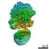
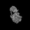
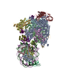
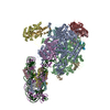
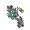
Journal: J Biol Chem / Year: 2023 Title: Cryo-EM structures of RNA polymerase II-nucleosome complexes rewrapping transcribed DNA. Authors: Munetaka Akatsu / Haruhiko Ehara / Tomoya Kujirai / Risa Fujita / Tomoko Ito / Ken Osumi / Mitsuo Ogasawara / Yoshimasa Takizawa / Shun-Ichi Sekine / Hitoshi Kurumizaka / Abstract: RNA polymerase II (RNAPII) transcribes DNA wrapped in the nucleosome by stepwise pausing, especially at nucleosomal superhelical locations -5 and -1 [SHL(-5) and SHL(-1), respectively]. In the ...RNA polymerase II (RNAPII) transcribes DNA wrapped in the nucleosome by stepwise pausing, especially at nucleosomal superhelical locations -5 and -1 [SHL(-5) and SHL(-1), respectively]. In the present study, we performed cryo-electron microscopy analyses of RNAPII-nucleosome complexes paused at a major nucleosomal pausing site, SHL(-1). We determined two previously undetected structures, in which the transcribed DNA behind RNAPII is sharply kinked at the RNAPII exit tunnel and rewrapped around the nucleosomal histones in front of RNAPII by DNA looping. This DNA kink shifts the DNA orientation toward the nucleosome, and the transcribed DNA region interacts with basic amino acid residues of histones H2A, H2B, and H3 exposed by the RNAPII-mediated nucleosomal DNA peeling. The DNA loop structure was not observed in the presence of the transcription elongation factors Spt4/5 and Elf1. These RNAPII-nucleosome structures provide important information for understanding the functional relevance of DNA looping during transcription elongation in the nucleosome.
In the structure databanks used in Yorodumi, some data are registered as the other names, "COVID-19 virus" and "2019-nCoV". Here are the details of the virus and the list of structure data.
Jan 31, 2019. EMDB accession codes are about to change! (news from PDBe EMDB page)
EMDB accession codes are about to change! (news from PDBe EMDB page)
The allocation of 4 digits for EMDB accession codes will soon come to an end. Whilst these codes will remain in use, new EMDB accession codes will include an additional digit and will expand incrementally as the available range of codes is exhausted. The current 4-digit format prefixed with “EMD-” (i.e. EMD-XXXX) will advance to a 5-digit format (i.e. EMD-XXXXX), and so on. It is currently estimated that the 4-digit codes will be depleted around Spring 2019, at which point the 5-digit format will come into force.
The EM Navigator/Yorodumi systems omit the EMD- prefix.
Related info.:Q: What is EMD? / ID/Accession-code notation in Yorodumi/EM Navigator
Yorodumi is a browser for structure data from EMDB, PDB, SASBDB, etc.
This page is also the successor to EM Navigator detail page, and also detail information page/front-end page for Omokage search.
The word "yorodu" (or yorozu) is an old Japanese word meaning "ten thousand". "mi" (miru) is to see.
Related info.:EMDB / PDB / SASBDB / Comparison of 3 databanks / Yorodumi Search / Aug 31, 2016. New EM Navigator & Yorodumi / Yorodumi Papers / Jmol/JSmol / Function and homology information / Changes in new EM Navigator and Yorodumi
 Movie
Movie Controller

 Yorodumi
 Open data
 Basic information
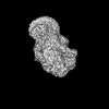
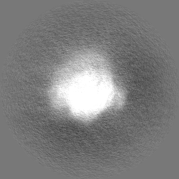
 Map data
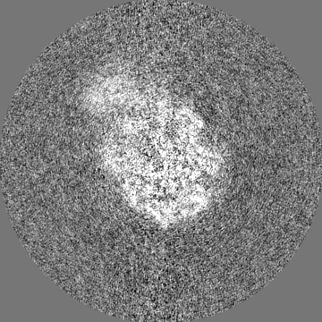
 Sample
 Keywords
 Komagataella phaffii (fungus) /
  Homo sapiens (human) / synthetic construct (others)
 Authors
 Japan, 10 items
  Citation
 Structure visualization
 Downloads & links
 EMDB map data format
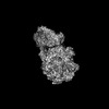
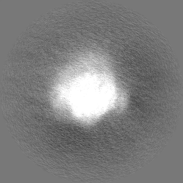
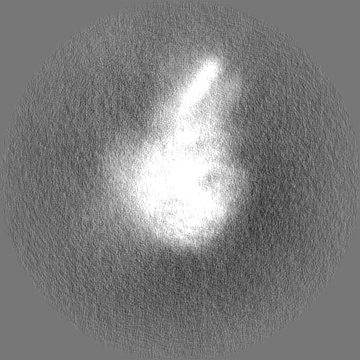
 emd_37848.png
 http://ftp.pdbj.org/pub/emdb/structures/EMD-37848
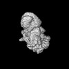

 Z (Sec.)
 Y (Row.)
 X (Col.)


 Sample components
 Processing
 Electron microscopy
 FIELD EMISSION GUN