 Movie
Movie Controller

[English] 日本語
 Yorodumi
- EMDB-37693: Cryo-EM structure of the 10-subunits Mmp1 complex from Mycobacter... -
+ Open data

- Basic information
| Entry |  | |||||||||||||||
|---|---|---|---|---|---|---|---|---|---|---|---|---|---|---|---|---|
| Title | Cryo-EM structure of the 10-subunits Mmp1 complex from Mycobacterium smegmatis | |||||||||||||||
 Map data Map data | ||||||||||||||||
 Sample Sample |
| |||||||||||||||
 Keywords Keywords | 10-subunits Mmp1 complex / STRUCTURAL PROTEIN | |||||||||||||||
| Function / homology | : / Type 2A encapsulin shell protein SrpI-like / Type 2A encapsulin shell protein SrpI-like / Major membrane protein I Function and homology information Function and homology information | |||||||||||||||
| Biological species |  Mycolicibacterium smegmatis (bacteria) Mycolicibacterium smegmatis (bacteria) | |||||||||||||||
| Method | single particle reconstruction / cryo EM / Resolution: 2.69 Å | |||||||||||||||
 Authors Authors | Zhang M / Tang Y / Gao Y / Liu X / Lan W / Liu Y / Ma M | |||||||||||||||
| Funding support |  China, 4 items China, 4 items
| |||||||||||||||
 Citation Citation | Journal: Commun Biol / Year: 2024 Title: The structural and functional analysis of mycobacteria cysteine desulfurase-loaded encapsulin. Authors: Yanting Tang / Yanyan Liu / Mingjing Zhang / Weiqi Lan / Mengyuan Ma / Cheng Chen / Saibin Wu / Rong Chen / Yiran Yan / Lu Feng / Ying Li / Luke W Guddat / Yan Gao / Xiang Liu / Zihe Rao /  Abstract: Encapsulin nanocompartments loaded with dedicated cargo proteins via unique targeting peptides, play a key role in stress resistance, iron storage and natural product biosynthesis. Mmp1 and cysteine ...Encapsulin nanocompartments loaded with dedicated cargo proteins via unique targeting peptides, play a key role in stress resistance, iron storage and natural product biosynthesis. Mmp1 and cysteine desulfurase (Enc-CD) have been identified as the most abundant representatives of family 2 encapsulin systems. However, the molecular assembly, catalytic mechanism, and physiological functions of the Mmp1 encapsulin system have not been studied in detail. Here we isolate and characterize an Enc-CD-loaded Mmp1 encapsulin system from Mycobacterium smegmatis mc155. The cryo-EM structure of the Mmp1 encapsulin and the crystal structure of the naked cargo Enc-CD have been determined. The structure shows that the Mmp1 protomer assembles two conformation models, the icosahedron (T = 1) and homodecamer, with the resolution of 2.60 Å and 2.69 Å. The Enc-CD at 2.10 Å resolution is dimeric and loaded into the Mmp1 (T = 1) encapsulin through the N-terminal long disordered region. Mmp1 encapsulin protects Enc-CD against oxidation as well as to maintain structural stability. These studies provide new insights into the mechanism by which Enc-CD-loaded encapsulin stores sulfur and provides a framework for discovery of new anti-mycobacterial therapeutics. | |||||||||||||||
| History |
|
- Structure visualization
| Supplemental images |
|---|

- Downloads & links
-EMDB archive
| Map data | emd_37693.map.gz | 117.9 MB | EMDB map data format | |
|---|---|---|---|---|
| Header (meta data) | emd-37693-v30.xmlemd-37693.xml | 16.9 KB 16.9 KB | Display Display | EMDB header |
| Images |  emd_37693.png emd_37693.png | 37.2 KB | ||
| Filedesc metadata | emd-37693.cif.gz | 5.8 KB | ||
| Others | emd_37693_half_map_1.map.gzemd_37693_half_map_2.map.gz | 116 MB 116 MB | ||
| Archive directory |  http://ftp.pdbj.org/pub/emdb/structures/EMD-37693ftp://ftp.pdbj.org/pub/emdb/structures/EMD-37693 http://ftp.pdbj.org/pub/emdb/structures/EMD-37693ftp://ftp.pdbj.org/pub/emdb/structures/EMD-37693 | HTTPS FTP |
-Validation report
| Summary document | emd_37693_validation.pdf.gz | 895.9 KB | Display | EMDB validaton report |
|---|---|---|---|---|
| Full document | emd_37693_full_validation.pdf.gz | 895.4 KB | Display | |
| Data in XML | emd_37693_validation.xml.gz | 14.1 KB | Display | |
| Data in CIF | emd_37693_validation.cif.gz | 16.4 KB | Display | |
| Arichive directory | https://ftp.pdbj.org/pub/emdb/validation_reports/EMD-37693ftp://ftp.pdbj.org/pub/emdb/validation_reports/EMD-37693 | HTTPS FTP |
-Related structure data
| Related structure data | 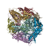 8wonMC  8wolC M: atomic model generated by this map C: citing same article ( |
|---|---|
| Similar structure data |


-Links
| EMDB pages | EMDB (EBI/PDBe) / EMDataResource |
|---|
-Map
| File | Download / File: emd_37693.map.gz / Format: CCP4 / Size: 125 MB / Type: IMAGE STORED AS FLOATING POINT NUMBER (4 BYTES) | ||||||||||||||||||||||||||||||||||||
|---|---|---|---|---|---|---|---|---|---|---|---|---|---|---|---|---|---|---|---|---|---|---|---|---|---|---|---|---|---|---|---|---|---|---|---|---|---|
| Projections & slices | Image control
Images are generated by Spider. | ||||||||||||||||||||||||||||||||||||
| Voxel size | X=Y=Z: 0.832 Å | ||||||||||||||||||||||||||||||||||||
| Density |
| ||||||||||||||||||||||||||||||||||||
| Symmetry | Space group: 1 | ||||||||||||||||||||||||||||||||||||
| Details | EMDB XML:
|
 Z (Sec.)
 Y (Row.)
 X (Col.)


-Supplemental data
-Half map: #2
| File | emd_37693_half_map_1.map | ||||||||||||
|---|---|---|---|---|---|---|---|---|---|---|---|---|---|
| Projections & Slices |
| ||||||||||||
| Density Histograms |


-Half map: #1
| File | emd_37693_half_map_2.map | ||||||||||||
|---|---|---|---|---|---|---|---|---|---|---|---|---|---|
| Projections & Slices |
| ||||||||||||
| Density Histograms |


- Sample components
-Entire : Mmp1 encapasulin
| Entire | Name: Mmp1 encapasulin |
|---|---|
| Components |
|
-Supramolecule #1: Mmp1 encapasulin
| Supramolecule | Name: Mmp1 encapasulin / type: complex / ID: 1 / Parent: 0 / Macromolecule list: all |
|---|---|
| Source (natural) | Organism: Mycolicibacterium smegmatis (bacteria) |
-Macromolecule #1: Major membrane protein I
| Macromolecule | Name: Major membrane protein I / type: protein_or_peptide / ID: 1 / Number of copies: 10 / Enantiomer: LEVO |
|---|---|
| Source (natural) | Organism: Mycolicibacterium smegmatis (bacteria) |
| Molecular weight | Theoretical: 31.769803 KDa |
| Recombinant expression | Organism: Mycolicibacterium smegmatis (bacteria) |
| Sequence | String: ANATKTVPQL STITPRFLLH LLSWVPVEAG IYRVNRVVNP DRVAIHSEAG AGTEEPLPET YVDYETHPRE YTLRSISTLL DVHTRVSDL YSSPHDQVTQ QLRLTIETIK ERQEYELVNN PEYGLLAQAT PEQTIQTLAG APTPDDLDAL ITKVWKTPAF F LTHPLGVA ...String: ANATKTVPQL STITPRFLLH LLSWVPVEAG IYRVNRVVNP DRVAIHSEAG AGTEEPLPET YVDYETHPRE YTLRSISTLL DVHTRVSDL YSSPHDQVTQ QLRLTIETIK ERQEYELVNN PEYGLLAQAT PEQTIQTLAG APTPDDLDAL ITKVWKTPAF F LTHPLGVA AFGRECTYRG VPPPTVSMYG AQFITWRGIP IVPSDKVPVE DGTTKFVLVR TGEERQGVVG LFQPGLVGEQ AP GLSVRFT GINRSAIASY LVTLYTSLAV LTDDALAVLD GVAVDQFHEY Q UniProtKB: Major membrane protein I |
-Experimental details
-Structure determination
| Method | cryo EM |
|---|---|
 Processing Processing | single particle reconstruction |
| Aggregation state | particle |
-Sample preparation
| Buffer | pH: 7.4 |
|---|---|
| Vitrification | Cryogen name: ETHANE |
- Electron microscopy
| Microscope | TFS KRIOS |
|---|---|
| Image recording | Film or detector model: GATAN K3 (6k x 4k) / Average electron dose: 60.0 e/Å2 |
| Electron beam | Acceleration voltage: 300 kV / Electron source:  FIELD EMISSION GUN FIELD EMISSION GUN |
| Electron optics | Illumination mode: FLOOD BEAM / Imaging mode: BRIGHT FIELD / Nominal defocus max: 1.8 µm / Nominal defocus min: 1.2 µm |
| Experimental equipment |  Model: Titan Krios / Image courtesy: FEI Company |
