 Movie
Movie Controller

[English] 日本語
 Yorodumi
- EMDB-37440: Cryo-EM structure of the inhibitor-bound Vo complex from Enteroco... -
+ Open data

- Basic information
| Entry |  | |||||||||
|---|---|---|---|---|---|---|---|---|---|---|
| Title | Cryo-EM structure of the inhibitor-bound Vo complex from Enterococcus hirae | |||||||||
 Map data Map data | ||||||||||
 Sample Sample |
| |||||||||
 Keywords Keywords | V-ATPase / Na+-transporting / membrane protein / ATP hydrolyses / HYDROLASE / HYDROLASE-HYDROLASE INHIBITOR complex | |||||||||
| Function / homology |  Function and homology information Function and homology informationproton-transporting V-type ATPase, V0 domain / vacuolar proton-transporting V-type ATPase complex / vacuolar acidification / sodium ion transport / proton-transporting ATPase activity, rotational mechanism / ATPase binding / identical protein binding / plasma membrane Similarity search - Function | |||||||||
| Biological species |  Enterococcus hirae ATCC 9790 (bacteria) Enterococcus hirae ATCC 9790 (bacteria) | |||||||||
| Method | single particle reconstruction / cryo EM / Resolution: 2.2 Å | |||||||||
 Authors Authors | Suzuki K / Mikuriya S / Adachi N / Kawasaki M / Senda T / Moriya T / Murata T | |||||||||
| Funding support |  Japan, 2 items Japan, 2 items
| |||||||||
 Citation Citation | Journal: Nat Struct Mol Biol / Year: 2025 Title: Na-V-ATPase inhibitor curbs VRE growth and unveils Na pathway structure. Authors: Kano Suzuki / Yoshiyuki Goto / Akihiro Otomo / Kouki Shimizu / Shohei Abe / Katsuhiko Moriyama / Satoshi Yasuda / Yusuke Hashimoto / Jun Kurushima / Sho Mikuriya / Fabiana L Imai / Naruhiko ...Authors: Kano Suzuki / Yoshiyuki Goto / Akihiro Otomo / Kouki Shimizu / Shohei Abe / Katsuhiko Moriyama / Satoshi Yasuda / Yusuke Hashimoto / Jun Kurushima / Sho Mikuriya / Fabiana L Imai / Naruhiko Adachi / Masato Kawasaki / Yumi Sato / Satoshi Ogasawara / So Iwata / Toshiya Senda / Mitsunori Ikeguchi / Haruyoshi Tomita / Ryota Iino / Toshio Moriya / Takeshi Murata / Abstract: Vancomycin-resistant Enterococcus faecium (VRE) is a major cause of nosocomial infections, particularly endocarditis and sepsis. With the diminishing effectiveness of antibiotics against VRE, new ...Vancomycin-resistant Enterococcus faecium (VRE) is a major cause of nosocomial infections, particularly endocarditis and sepsis. With the diminishing effectiveness of antibiotics against VRE, new antimicrobial agents are urgently needed. Our previous research demonstrated the crucial role of Na-transporting V-ATPase in Enterococcus hirae for growth under alkaline conditions. In this study, we identified a compound, V-161, from 70,600 compounds, which markedly inhibits E. hirae V-ATPase activity. V-161 not only inhibits VRE growth in alkaline conditions but also significantly suppresses VRE colonization in the mouse small intestine. Furthermore, we unveiled the high-resolution structure of the membrane V part due to V-161 binding. V-161 binds to the interface of the c-ring and a-subunit, constituting the Na transport pathway in the membrane, thereby halting its rotation. This structural insight presents potential avenues for developing therapeutic agents for VRE treatment and elucidates the Na transport pathway and mechanism. | |||||||||
| History |
|
- Structure visualization
| Supplemental images |
|---|

- Downloads & links
-EMDB archive
| Map data | emd_37440.map.gz | 479.4 MB | EMDB map data format | |
|---|---|---|---|---|
| Header (meta data) | emd-37440-v30.xmlemd-37440.xml | 23.3 KB 23.3 KB | Display Display | EMDB header |
| FSC (resolution estimation) | emd_37440_fsc.xml | 18 KB | Display | FSC data file |
| Images |  emd_37440.png emd_37440.png | 152.8 KB | ||
| Masks | emd_37440_msk_1.map | 512 MB | Mask map | |
| Filedesc metadata | emd-37440.cif.gz | 7.4 KB | ||
| Others | emd_37440_half_map_1.map.gzemd_37440_half_map_2.map.gz | 410.9 MB 410.7 MB | ||
| Archive directory |  http://ftp.pdbj.org/pub/emdb/structures/EMD-37440ftp://ftp.pdbj.org/pub/emdb/structures/EMD-37440 http://ftp.pdbj.org/pub/emdb/structures/EMD-37440ftp://ftp.pdbj.org/pub/emdb/structures/EMD-37440 | HTTPS FTP |
-Related structure data
| Related structure data | 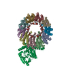 8wciMC M: atomic model generated by this map C: citing same article ( |
|---|---|
| Similar structure data |
-Links
| EMDB pages | EMDB (EBI/PDBe) / EMDataResource |
|---|---|
| Related items in Molecule of the Month |


-Map
| File | Download / File: emd_37440.map.gz / Format: CCP4 / Size: 512 MB / Type: IMAGE STORED AS FLOATING POINT NUMBER (4 BYTES) | ||||||||||||||||||||||||||||||||||||
|---|---|---|---|---|---|---|---|---|---|---|---|---|---|---|---|---|---|---|---|---|---|---|---|---|---|---|---|---|---|---|---|---|---|---|---|---|---|
| Projections & slices | Image control
Images are generated by Spider. | ||||||||||||||||||||||||||||||||||||
| Voxel size | X=Y=Z: 0.83 Å | ||||||||||||||||||||||||||||||||||||
| Density |
| ||||||||||||||||||||||||||||||||||||
| Symmetry | Space group: 1 | ||||||||||||||||||||||||||||||||||||
| Details | EMDB XML:
|
 Z (Sec.)
 Y (Row.)
 X (Col.)


-Supplemental data
-Mask #1
| File | emd_37440_msk_1.map | ||||||||||||
|---|---|---|---|---|---|---|---|---|---|---|---|---|---|
| Projections & Slices |
| ||||||||||||
| Density Histograms |


-Half map: #1
| File | emd_37440_half_map_1.map | ||||||||||||
|---|---|---|---|---|---|---|---|---|---|---|---|---|---|
| Projections & Slices |
| ||||||||||||
| Density Histograms |


-Half map: #2
| File | emd_37440_half_map_2.map | ||||||||||||
|---|---|---|---|---|---|---|---|---|---|---|---|---|---|
| Projections & Slices |
| ||||||||||||
| Density Histograms |


- Sample components
-Entire : V-ATPase
| Entire | Name: V-ATPase |
|---|---|
| Components |
|
-Supramolecule #1: V-ATPase
| Supramolecule | Name: V-ATPase / type: complex / ID: 1 / Parent: 0 / Macromolecule list: #1-#2 |
|---|---|
| Source (natural) | Organism: Enterococcus hirae ATCC 9790 (bacteria) |
| Molecular weight | Theoretical: 735 KDa |
-Macromolecule #1: V-type sodium ATPase subunit K
| Macromolecule | Name: V-type sodium ATPase subunit K / type: protein_or_peptide / ID: 1 / Number of copies: 10 / Enantiomer: LEVO |
|---|---|
| Source (natural) | Organism: Enterococcus hirae ATCC 9790 (bacteria) |
| Molecular weight | Theoretical: 16.043918 KDa |
| Recombinant expression | Organism: |
| Sequence | String: MMDYLITQNG GMVFAVLAMA TATIFSGIGS AKGVGMTGEA AAALTTSQPE KFGQALILQL LPGTQGLYGF VIAFLIFINL GSDMSVVQG LNFLGASLPI AFTGLFSGIA QGKVAAAGIQ ILAKKPEHAT KGIIFAAMVE TYAILGFVIS FLLVLNA UniProtKB: V-type sodium ATPase subunit K |
-Macromolecule #2: V-type sodium ATPase subunit I
| Macromolecule | Name: V-type sodium ATPase subunit I / type: protein_or_peptide / ID: 2 / Number of copies: 1 / Enantiomer: LEVO |
|---|---|
| Source (natural) | Organism: Enterococcus hirae ATCC 9790 (bacteria) |
| Molecular weight | Theoretical: 76.5155 KDa |
| Recombinant expression | Organism: |
| Sequence | String: MAVTKMEKVT LISDKKNREI LLQAVQGLHA VEIRDLFQES ENNQWVETFF PEPEMIDKDK ELAKLSYKLT DIRTAIQFIE HHGEKSQKK QHLKRRELSL DTLEKNYSEE AFSKKLEEVL LLKEQWEQLV DERQQLEDQE NWLLNWQNLD LAPKAFDSQM T KLVIGTVN ...String: MAVTKMEKVT LISDKKNREI LLQAVQGLHA VEIRDLFQES ENNQWVETFF PEPEMIDKDK ELAKLSYKLT DIRTAIQFIE HHGEKSQKK QHLKRRELSL DTLEKNYSEE AFSKKLEEVL LLKEQWEQLV DERQQLEDQE NWLLNWQNLD LAPKAFDSQM T KLVIGTVN AKNAESFKAE VAEINEAYLE EINSSPTTTY FAYIVLRADE SRMEEIASRY GFVKEDYLYE GTPQQQLVAA KQ SLQEIKD QQKKLSSAIG ACSGYIKDFE WTEEIFLARS EREAIKDRII HTPYLILIQG WVDHEEKQEL IHMLQNILAS EEV YLTFDE PTDNEIAEEV PTKLKNHPIV APFEMLTEMY SLPKYEEVDP TPWMMPFYLV FFGMMVADIG YGLLMFLGAF LLQK LVVLP RGMQRFAKFF EILAIPSIIW GFIYSSFFGA ALPKEIFGIH LPFPILSTTD DVNTILILSV IFGLIQILVG LFIAA KEHI KRKAYVDAVN DGFAWQGILL GIILILLGTM ILKNNAFVYL GGALAVLSAV CILIIPVFQS SSKAKGIAKG AYNLYG LTG YIGDLVSYTR LMALGISGGS IAAAFNMLVA FMPPAARFSV GILLIIVLQA LNMFLTLLSA YVHGARLQYV EFFGKFY TG GGRSFKPLKT VEKYVNINHK KKEHLYFQGG UniProtKB: V-type sodium ATPase subunit I |
-Macromolecule #3: CARDIOLIPIN
| Macromolecule | Name: CARDIOLIPIN / type: ligand / ID: 3 / Number of copies: 5 / Formula: CDL |
|---|---|
| Molecular weight | Theoretical: 1.464043 KDa |
| Chemical component information |  ChemComp-CDL: |
-Macromolecule #4: SODIUM ION
| Macromolecule | Name: SODIUM ION / type: ligand / ID: 4 / Number of copies: 9 |
|---|---|
| Molecular weight | Theoretical: 22.99 Da |
-Macromolecule #5: N,N-dimethyl-4-(5-methyl-1H-benzimidazol-2-yl)aniline
| Macromolecule | Name: N,N-dimethyl-4-(5-methyl-1H-benzimidazol-2-yl)aniline / type: ligand / ID: 5 / Number of copies: 1 / Formula: W3K |
|---|---|
| Molecular weight | Theoretical: 251.326 Da |
-Macromolecule #6: water
| Macromolecule | Name: water / type: ligand / ID: 6 / Number of copies: 224 / Formula: HOH |
|---|---|
| Molecular weight | Theoretical: 18.015 Da |
| Chemical component information |  ChemComp-HOH: |
-Experimental details
-Structure determination
| Method | cryo EM |
|---|---|
 Processing Processing | single particle reconstruction |
| Aggregation state | particle |
-Sample preparation
| Concentration | 6 mg/mL | |||||||||||||||
|---|---|---|---|---|---|---|---|---|---|---|---|---|---|---|---|---|
| Buffer | pH: 7.5 Component:
| |||||||||||||||
| Grid | Model: Quantifoil R1.2/1.3 / Material: COPPER / Mesh: 300 | |||||||||||||||
| Vitrification | Cryogen name: ETHANE / Chamber humidity: 100 % / Chamber temperature: 277 K / Instrument: FEI VITROBOT MARK IV |
- Electron microscopy
| Microscope | TFS KRIOS |
|---|---|
| Specialist optics | Energy filter - Name: GIF Bioquantum / Energy filter - Slit width: 15 eV |
| Image recording | Film or detector model: GATAN K3 BIOQUANTUM (6k x 4k) / Average electron dose: 49.0 e/Å2 |
| Electron beam | Acceleration voltage: 300 kV / Electron source:  FIELD EMISSION GUN FIELD EMISSION GUN |
| Electron optics | Illumination mode: FLOOD BEAM / Imaging mode: BRIGHT FIELD / Nominal defocus max: 2.5 µm / Nominal defocus min: 1.0 µm |
| Experimental equipment |  Model: Titan Krios / Image courtesy: FEI Company |


