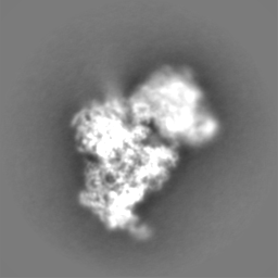
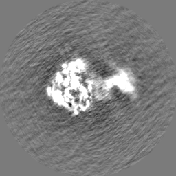
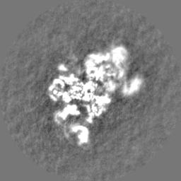
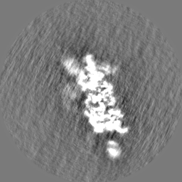
- EMDB-36854: Cryo-EM structure of 30S ribosome with cleaved AP-mRNA bound complex I -
+
データを開く
IDまたはキーワード:
読み込み中...
-
基本情報
登録情報
データベース: EMDB / ID: EMD-36854
タイトル
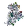
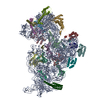
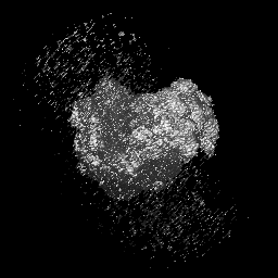
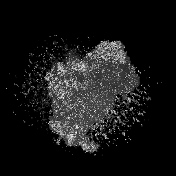
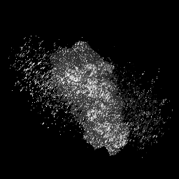
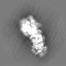
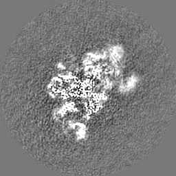
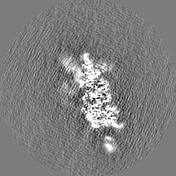
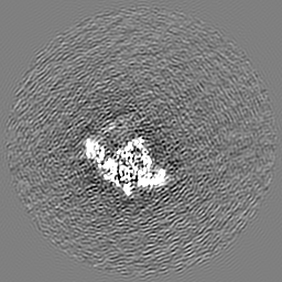
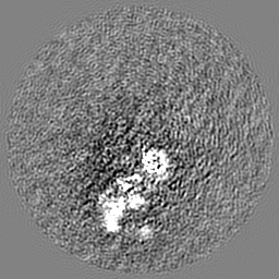
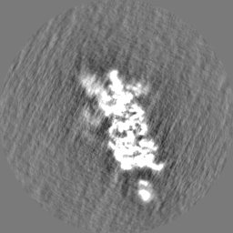
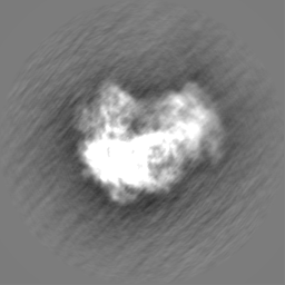
Cryo-EM structure of 30S ribosome with cleaved AP-mRNA bound complex I
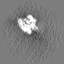
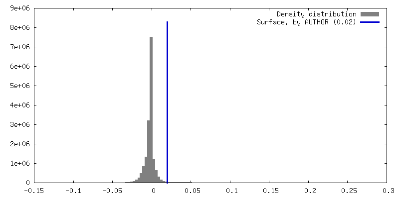
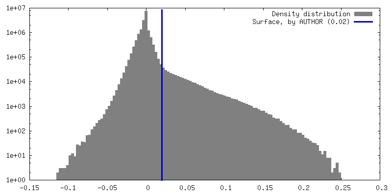
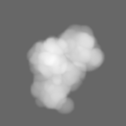
マップデータ
試料
複合体: 30S ribosome with cleaved AP-mRNA
タンパク質・ペプチド: x 20種
RNA: x 2種
キーワード
Ribosome / AP-mRNA
機能・相同性
機能・相同性情報
ornithine decarboxylase inhibitor activity / transcription antitermination factor activity, RNA binding / misfolded RNA binding / Group I intron splicing / RNA folding / four-way junction DNA binding / negative regulation of translational initiation / regulation of mRNA stability / mRNA regulatory element binding translation repressor activity / positive regulation of RNA splicing ...ornithine decarboxylase inhibitor activity / transcription antitermination factor activity, RNA binding / misfolded RNA binding / Group I intron splicing / RNA folding / four-way junction DNA binding / negative regulation of translational initiation / regulation of mRNA stability / mRNA regulatory element binding translation repressor activity / positive regulation of RNA splicing / transcription elongation factor complex / regulation of DNA-templated transcription elongation / DNA endonuclease activity / transcription antitermination / DNA-templated transcription termination / maintenance of translational fidelity / mRNA 5'-UTR binding / regulation of translation / ribosome biogenesis / ribosomal small subunit biogenesis / ribosomal small subunit assembly / small ribosomal subunit / cytosolic small ribosomal subunit / small ribosomal subunit rRNA binding / cytoplasmic translation / tRNA binding / negative regulation of translation / rRNA binding / ribosome / structural constituent of ribosome / translation / response to antibiotic / mRNA binding / RNA binding / zinc ion binding / membrane / cytosol / cytoplasm 類似検索 - 分子機能
Ribosomal protein S21, conserved site / Ribosomal protein S21 signature. / Ribosomal protein S14, bacterial/plastid / Ribosomal protein S21 superfamily / Ribosomal protein S16, conserved site / Ribosomal protein S16 signature. / Ribosomal protein S21 / Ribosomal protein S21 / Ribosomal protein S19, bacterial-type / Ribosomal protein S3, bacterial-type ...Ribosomal protein S21, conserved site / Ribosomal protein S21 signature. / Ribosomal protein S14, bacterial/plastid / Ribosomal protein S21 superfamily / Ribosomal protein S16, conserved site / Ribosomal protein S16 signature. / Ribosomal protein S21 / Ribosomal protein S21 / Ribosomal protein S19, bacterial-type / Ribosomal protein S3, bacterial-type / Ribosomal protein S13, bacterial-type / Ribosomal protein S6, conserved site / Ribosomal protein S6 signature. / Ribosomal protein S7, bacterial/organellar-type / Ribosomal protein S9, bacterial/plastid / Ribosomal protein S11, bacterial-type / Ribosomal protein S20 / Ribosomal protein S20 superfamily / Ribosomal protein S20 / Ribosomal protein S4, bacterial-type / 30S ribosomal protein S17 / Ribosomal protein S5, bacterial-type / Ribosomal protein S2, bacteria/mitochondria/plastid / Ribosomal protein S18, conserved site / Ribosomal protein S18 signature. / Ribosomal protein S6, plastid/chloroplast / Ribosomal protein S16 / Ribosomal protein S16 domain superfamily / Ribosomal protein S16 / Ribosomal protein S15, bacterial-type / Ribosomal protein S12, bacterial-type / Ribosomal protein S18 / Ribosomal protein S18 / Ribosomal protein S18 superfamily / K Homology domain / K homology RNA-binding domain / Ribosomal protein S6 / Ribosomal protein S6 / Ribosomal protein S6 superfamily / Ribosomal protein S2 signature 2. / Translation elongation factor EF1B/ribosomal protein S6 / Ribosomal protein S3, conserved site / Ribosomal protein S3 signature. / Ribosomal protein S10, conserved site / Ribosomal protein S10 signature. / Ribosomal protein S14, conserved site / Ribosomal protein S14 signature. / Ribosomal protein S2 signature 1. / KH domain / Type-2 KH domain profile. / K Homology domain, type 2 / Ribosomal protein S3, C-terminal / Ribosomal protein S3, C-terminal domain / Ribosomal protein S3, C-terminal domain superfamily / Ribosomal protein S15/S19, conserved site / Ribosomal protein S19 signature. / Ribosomal protein S10 / Ribosomal protein S19/S15 / Ribosomal protein S19/S15, superfamily / Ribosomal protein S19 / : / Ribosomal protein S5, N-terminal, conserved site / Ribosomal protein S5 signature. / Ribosomal protein S7, conserved site / Ribosomal protein S2, conserved site / Ribosomal protein S7 signature. / Ribosomal protein S2 / Ribosomal protein S2, flavodoxin-like domain superfamily / Ribosomal protein S2 / Ribosomal protein S17, conserved site / Ribosomal protein S17 signature. / K homology domain superfamily, prokaryotic type / Ribosomal protein S5 / Ribosomal protein S13, conserved site / Ribosomal protein S13 signature. / S5 double stranded RNA-binding domain profile. / Ribosomal protein S5, N-terminal / Ribosomal protein S13 / 30s ribosomal protein S13, C-terminal / Ribosomal protein S13/S18 / Ribosomal protein S5, C-terminal / Ribosomal protein S13 family profile. / Ribosomal protein S5, N-terminal domain / Ribosomal protein S5, C-terminal domain / Ribosomal protein S8 signature. / Ribosomal protein S4/S9 N-terminal domain / Ribosomal protein S4, conserved site / Ribosomal protein S15 signature. / Ribosomal protein S4 signature. / Ribosomal protein S4/S9 N-terminal domain / Ribosomal protein S4/S9, N-terminal / Ribosomal protein S14 / Ribosomal protein S14p/S29e / Ribosomal protein S4/S9 / K homology domain-like, alpha/beta / Ribosomal protein S8 / Ribosomal protein S8 superfamily / Ribosomal protein S8 / S4 RNA-binding domain profile. / Ribosomal protein S13-like, H2TH 類似検索 - ドメイン・相同性
Small ribosomal subunit protein bS6 / Small ribosomal subunit protein uS7 / Small ribosomal subunit protein uS10 / Small ribosomal subunit protein uS11 / Small ribosomal subunit protein uS12 / Small ribosomal subunit protein uS13 / Small ribosomal subunit protein bS16 / Small ribosomal subunit protein bS18 / Small ribosomal subunit protein uS19 / Small ribosomal subunit protein bS20 ...Small ribosomal subunit protein bS6 / Small ribosomal subunit protein uS7 / Small ribosomal subunit protein uS10 / Small ribosomal subunit protein uS11 / Small ribosomal subunit protein uS12 / Small ribosomal subunit protein uS13 / Small ribosomal subunit protein bS16 / Small ribosomal subunit protein bS18 / Small ribosomal subunit protein uS19 / Small ribosomal subunit protein bS20 / Small ribosomal subunit protein uS2 / Small ribosomal subunit protein uS3 / Small ribosomal subunit protein uS4 / Small ribosomal subunit protein uS5 / Small ribosomal subunit protein uS8 / Small ribosomal subunit protein uS9 / Small ribosomal subunit protein uS15 / Small ribosomal subunit protein uS14 / Small ribosomal subunit protein uS17 / Small ribosomal subunit protein bS21 類似検索 - 構成要素
Council of Scientific & Industrial Research (CSIR)
CII7032 & MLP0107
インド
Department of Biotechnology (DBT, India)
GAP0384
インド
引用
ジャーナル: Nucleic Acids Res / 年: 2024 タイトル: Bacterial Rps3 counters oxidative and UV stress by recognizing and processing AP-sites on mRNA via a novel mechanism. 著者: Mohammad Afsar / Ankita Shukla / Faiz Ali / Rahul Kumar Maurya / Suman Bharti / Nelam Kumar / Mohammad Sadik / Surabhi Chandra / Huma Rahil / Sanjay Kumar / Imran Ansari / Farheen Jahan / ...著者: Mohammad Afsar / Ankita Shukla / Faiz Ali / Rahul Kumar Maurya / Suman Bharti / Nelam Kumar / Mohammad Sadik / Surabhi Chandra / Huma Rahil / Sanjay Kumar / Imran Ansari / Farheen Jahan / Saman Habib / Tanweer Hussain / Manju Yasoda Krishnan / Ravishankar Ramachandran / 要旨: Lesions and stable secondary structures in mRNA severely impact the translation efficiency, causing ribosome stalling and collisions. Prokaryotic ribosomal proteins Rps3, Rps4 and Rps5, located in ...Lesions and stable secondary structures in mRNA severely impact the translation efficiency, causing ribosome stalling and collisions. Prokaryotic ribosomal proteins Rps3, Rps4 and Rps5, located in the mRNA entry tunnel, form the mRNA helicase center and unwind stable mRNA secondary structures during translation. However, the mechanism underlying the detection of lesions on translating mRNA is unclear. We used Cryo-EM, biochemical assays, and knockdown experiments to investigate the apurinic/apyrimidinic (AP) endoribonuclease activity of bacterial ribosomes on AP-site containing mRNA. Our biochemical assays show that Rps3, specifically the 130RR131 motif, is important for recognizing and performing the AP-endoribonuclease activity. Furthermore, structural analysis revealed cleaved mRNA product in the 30S ribosome entry tunnel. Additionally, knockdown studies in Mycobacterium tuberculosis confirmed the protective role of Rps3 against oxidative and UV stress. Overall, our results show that prokaryotic Rps3 recognizes and processes AP-sites on mRNA via a novel mechanism that is distinct from eukaryotes.
 ムービー
ムービー コントローラー

 万見
万見 データを開く
 基本情報
基本情報
 マップデータ
 試料
試料 キーワード
キーワード 機能・相同性情報
 データ登録者
 インド, 3件
  引用
引用 構造の表示
構造の表示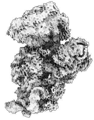
 ダウンロードとリンク
 emd_36854.png
 http://ftp.pdbj.org/pub/emdb/structures/EMD-36854


 Z (Sec.)
 Y (Row.)
 X (Col.)


 試料の構成要素
 解析
解析 電子顕微鏡法
 FIELD EMISSION GUN