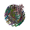
Journal: Biochim Biophys Acta Bioenerg / Year: 2024 Title: Molecular structure and characterization of the Thermochromatium tepidum light-harvesting 1 photocomplex produced in a foreign host. Authors: Yi-Hao Yan / Guang-Lei Wang / Xing-Yu Yue / Fei Ma / Michael T Madigan / Zheng-Yu Wang-Otomo / Mei-Juan Zou / Long-Jiang Yu / Abstract: Purple phototrophic bacteria possess light-harvesting 1 and reaction center (LH1-RC) core complexes that play a key role in converting solar energy to chemical energy. High-resolution structures of ...Purple phototrophic bacteria possess light-harvesting 1 and reaction center (LH1-RC) core complexes that play a key role in converting solar energy to chemical energy. High-resolution structures of LH1-RC and RC complexes have been intensively studied and have yielded critical insight into the architecture and interactions of their proteins, pigments, and cofactors. Nevertheless, a detailed picture of the structure and assembly of LH1-only complexes is lacking due to the intimate association between LH1 and the RC. To study the intrinsic properties and structure of an LH1-only complex, a genetic system was constructed to express the Thermochromatium (Tch.) tepidum LH1 complex heterologously in a modified Rhodospirillum rubrum mutant strain. The heterologously expressed Tch. tepidum LH1 complex was isolated in a pure form free of the RC and exhibited the characteristic absorption properties of Tch. tepidum. Cryo-EM structures of the LH1-only complexes revealed a closed circular ring consisting of either 14 or 15 αβ-subunits, making it the smallest completely closed LH1 complex discovered thus far. Surprisingly, the Tch. tepidum LH1-only complex displayed even higher thermostability than that of the native LH1-RC complex. These results reveal previously unsuspected plasticity of the LH1 complex, provide new insights into the structure and assembly of the LH1-RC complex, and show how molecular genetics can be exploited to study membrane proteins from phototrophic organisms whose genetic manipulation is not yet possible.
In the structure databanks used in Yorodumi, some data are registered as the other names, "COVID-19 virus" and "2019-nCoV". Here are the details of the virus and the list of structure data.
Jan 31, 2019. EMDB accession codes are about to change! (news from PDBe EMDB page)
EMDB accession codes are about to change! (news from PDBe EMDB page)
The allocation of 4 digits for EMDB accession codes will soon come to an end. Whilst these codes will remain in use, new EMDB accession codes will include an additional digit and will expand incrementally as the available range of codes is exhausted. The current 4-digit format prefixed with “EMD-” (i.e. EMD-XXXX) will advance to a 5-digit format (i.e. EMD-XXXXX), and so on. It is currently estimated that the 4-digit codes will be depleted around Spring 2019, at which point the 5-digit format will come into force.
The EM Navigator/Yorodumi systems omit the EMD- prefix.
Related info.:Q: What is EMD? / ID/Accession-code notation in Yorodumi/EM Navigator
Yorodumi is a browser for structure data from EMDB, PDB, SASBDB, etc.
This page is also the successor to EM Navigator detail page, and also detail information page/front-end page for Omokage search.
The word "yorodu" (or yorozu) is an old Japanese word meaning "ten thousand". "mi" (miru) is to see.
Related info.:EMDB / PDB / SASBDB / Comparison of 3 databanks / Yorodumi Search / Aug 31, 2016. New EM Navigator & Yorodumi / Yorodumi Papers / Jmol/JSmol / Function and homology information / Changes in new EM Navigator and Yorodumi
 Movie
Movie Controller

 Yorodumi
 Open data
 Basic information
 Map data
 Sample
 Keywords
 Function and homology information
 Thermochromatium tepidum (bacteria)
 Authors
 China, 2 items
  Citation

 Structure visualization
 Downloads & links
 emd_36154.png
 http://ftp.pdbj.org/pub/emdb/structures/EMD-36154
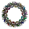

 Z (Sec.)
 Y (Row.)
 X (Col.)


 Sample components


 Processing
 Electron microscopy
 FIELD EMISSION GUN