Journal: Biochim Biophys Acta / Year: 2014 Title: Structures of mitochondrial oxidative phosphorylation supercomplexes and mechanisms for their stabilisation. Authors: Yuriy Chaban / Egbert J Boekema / Natalya V Dudkina / Abstract: Oxidative phosphorylation (OXPHOS) is the main source of energy in eukaryotic cells. This process is performed by means of electron flow between four enzymes, of which three are proton pumps, in the ...Oxidative phosphorylation (OXPHOS) is the main source of energy in eukaryotic cells. This process is performed by means of electron flow between four enzymes, of which three are proton pumps, in the inner mitochondrial membrane. The energy accumulated in the proton gradient over the inner membrane is utilized for ATP synthesis by a fifth OXPHOS complex, ATP synthase. Four of the OXPHOS protein complexes associate into stable entities called respiratory supercomplexes. This review summarises the current view on the arrangement of the electron transport chain in mitochondrial cristae. The functional role of the supramolecular organisation of the OXPHOS system and the factors that stabilise such organisation are highlighted. This article is part of a Special Issue entitled: Dynamic and ultrastructure of bioenergetic membranes and their components.
History
Deposition
Jan 27, 2017
-
Header (metadata) release
Feb 22, 2017
-
Map release
Mar 1, 2017
-
Update
Jul 26, 2017
-
Current status
Jul 26, 2017
Processing site: PDBe / Status: Released
-
Structure visualization
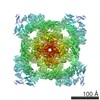
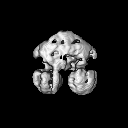
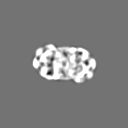
Movie
Surface view with section colored by density value
In the structure databanks used in Yorodumi, some data are registered as the other names, "COVID-19 virus" and "2019-nCoV". Here are the details of the virus and the list of structure data.
Jan 31, 2019. EMDB accession codes are about to change! (news from PDBe EMDB page)
EMDB accession codes are about to change! (news from PDBe EMDB page)
The allocation of 4 digits for EMDB accession codes will soon come to an end. Whilst these codes will remain in use, new EMDB accession codes will include an additional digit and will expand incrementally as the available range of codes is exhausted. The current 4-digit format prefixed with “EMD-” (i.e. EMD-XXXX) will advance to a 5-digit format (i.e. EMD-XXXXX), and so on. It is currently estimated that the 4-digit codes will be depleted around Spring 2019, at which point the 5-digit format will come into force.
The EM Navigator/Yorodumi systems omit the EMD- prefix.
Related info.:Q: What is EMD? / ID/Accession-code notation in Yorodumi/EM Navigator
Yorodumi is a browser for structure data from EMDB, PDB, SASBDB, etc.
This page is also the successor to EM Navigator detail page, and also detail information page/front-end page for Omokage search.
The word "yorodu" (or yorozu) is an old Japanese word meaning "ten thousand". "mi" (miru) is to see.
Related info.:EMDB / PDB / SASBDB / Comparison of 3 databanks / Yorodumi Search / Aug 31, 2016. New EM Navigator & Yorodumi / Yorodumi Papers / Jmol/JSmol / Function and homology information / Changes in new EM Navigator and Yorodumi
 Movie
Movie Controller


 Open data
 Basic information
 Map data
 Sample
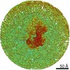
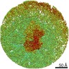
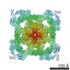
 Tetrahymena (eukaryote)
 Authors
 Citation

 Structure visualization
 Movie viewer
 UCSF Chimera
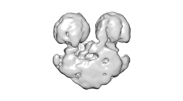
 Downloads & links
 emd_3582.png
 http://ftp.pdbj.org/pub/emdb/structures/EMD-3582
 Z (Sec.)
 Y (Row.)
 X (Col.)


 Sample components
 Processing
 Electron microscopy