[English] 日本語
 Yorodumi
- EMDB-35773: Human Consensus Olfactory Receptor OR52c in Complex with Octanoic... -
+ Open data
- Basic information
| Entry |  | |||||||||
|---|---|---|---|---|---|---|---|---|---|---|
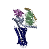
| Title | Human Consensus Olfactory Receptor OR52c in Complex with Octanoic Acid (OCA) and G Protein (G protein-focused map) | |||||||||
 Map data Map data | This is a G protein-focused map of human consensus olfactory receptor OR52c in complex with octanoic Acid (OCA) and G protein. This map was generated with Local refinement tool in cryoSPARC. | |||||||||
 Sample Sample |
| |||||||||
 Keywords Keywords | Olfactory Receptor / G Protein / MEMBRANE PROTEIN / GPCR / Olfactory GPCR | |||||||||
| Biological species |  Homo sapiens (human) Homo sapiens (human) | |||||||||
| Method | single particle reconstruction / cryo EM / Resolution: 2.84 Å | |||||||||
 Authors Authors | Choi CW / Bae J / Choi H-J | |||||||||
| Funding support |  Korea, Republic Of, 1 items Korea, Republic Of, 1 items
| |||||||||
 Citation Citation |  Journal: Nat Commun / Year: 2023 Journal: Nat Commun / Year: 2023Title: Understanding the molecular mechanisms of odorant binding and activation of the human OR52 family. Authors: Chulwon Choi / Jungnam Bae / Seonghan Kim / Seho Lee / Hyunook Kang / Jinuk Kim / Injin Bang / Kiheon Kim / Won-Ki Huh / Chaok Seok / Hahnbeom Park / Wonpil Im / Hee-Jung Choi /   Abstract: Structural and mechanistic studies on human odorant receptors (ORs), key in olfactory signaling, are challenging because of their low surface expression in heterologous cells. The recent structure of ...Structural and mechanistic studies on human odorant receptors (ORs), key in olfactory signaling, are challenging because of their low surface expression in heterologous cells. The recent structure of OR51E2 bound to propionate provided molecular insight into odorant recognition, but the lack of an inactive OR structure limited understanding of the activation mechanism of ORs upon odorant binding. Here, we determined the cryo-electron microscopy structures of consensus OR52 (OR52), a representative of the OR52 family, in the ligand-free (apo) and octanoate-bound states. The apo structure of OR52 reveals a large opening between transmembrane helices (TMs) 5 and 6. A comparison between the apo and active structures of OR52 demonstrates the inward and outward movements of the extracellular and intracellular segments of TM6, respectively. These results, combined with molecular dynamics simulations and signaling assays, shed light on the molecular mechanisms of odorant binding and activation of the OR52 family. | |||||||||
| History |
|
- Structure visualization
| Supplemental images |
|---|
- Downloads & links
-EMDB archive
| Map data |  emd_35773.map.gz emd_35773.map.gz | 230.3 MB |  EMDB map data format EMDB map data format | |
|---|---|---|---|---|
| Header (meta data) |  emd-35773-v30.xml emd-35773-v30.xml emd-35773.xml emd-35773.xml | 17.3 KB 17.3 KB | Display Display |  EMDB header EMDB header |
| FSC (resolution estimation) |  emd_35773_fsc.xml emd_35773_fsc.xml | 13.2 KB | Display |  FSC data file FSC data file |
| Images |  emd_35773.png emd_35773.png | 119 KB | ||
| Filedesc metadata |  emd-35773.cif.gz emd-35773.cif.gz | 4.7 KB | ||
| Others |  emd_35773_half_map_1.map.gz emd_35773_half_map_1.map.gz emd_35773_half_map_2.map.gz emd_35773_half_map_2.map.gz | 226.3 MB 226.3 MB | ||
| Archive directory |  http://ftp.pdbj.org/pub/emdb/structures/EMD-35773 http://ftp.pdbj.org/pub/emdb/structures/EMD-35773 ftp://ftp.pdbj.org/pub/emdb/structures/EMD-35773 ftp://ftp.pdbj.org/pub/emdb/structures/EMD-35773 | HTTPS FTP |
-Related structure data
- Links
Links
| EMDB pages |  EMDB (EBI/PDBe) / EMDB (EBI/PDBe) /  EMDataResource EMDataResource |
|---|
- Map
Map
| File |  Download / File: emd_35773.map.gz / Format: CCP4 / Size: 244.1 MB / Type: IMAGE STORED AS FLOATING POINT NUMBER (4 BYTES) Download / File: emd_35773.map.gz / Format: CCP4 / Size: 244.1 MB / Type: IMAGE STORED AS FLOATING POINT NUMBER (4 BYTES) | ||||||||||||||||||||||||||||||||||||
|---|---|---|---|---|---|---|---|---|---|---|---|---|---|---|---|---|---|---|---|---|---|---|---|---|---|---|---|---|---|---|---|---|---|---|---|---|---|
| Annotation | This is a G protein-focused map of human consensus olfactory receptor OR52c in complex with octanoic Acid (OCA) and G protein. This map was generated with Local refinement tool in cryoSPARC. | ||||||||||||||||||||||||||||||||||||
| Projections & slices | Image control
Images are generated by Spider. | ||||||||||||||||||||||||||||||||||||
| Voxel size | X=Y=Z: 0.851 Å | ||||||||||||||||||||||||||||||||||||
| Density |
| ||||||||||||||||||||||||||||||||||||
| Symmetry | Space group: 1 | ||||||||||||||||||||||||||||||||||||
| Details | EMDB XML:
|
-Supplemental data
-Half map: This is a half B map of the G protein-focused map.
| File | emd_35773_half_map_1.map | ||||||||||||
|---|---|---|---|---|---|---|---|---|---|---|---|---|---|
| Annotation | This is a half B map of the G protein-focused map. | ||||||||||||
| Projections & Slices |
| ||||||||||||
| Density Histograms |
-Half map: This is a half A map of the G protein-focused map.
| File | emd_35773_half_map_2.map | ||||||||||||
|---|---|---|---|---|---|---|---|---|---|---|---|---|---|
| Annotation | This is a half A map of the G protein-focused map. | ||||||||||||
| Projections & Slices |
| ||||||||||||
| Density Histograms |
- Sample components
-Entire : Complex of Consensus Olfactory receptor OR52c in complex with Oct...
| Entire | Name: Complex of Consensus Olfactory receptor OR52c in complex with Octanoic acid, G protein and Nanobody 35 (Nb35) |
|---|---|
| Components |
|
-Supramolecule #1: Complex of Consensus Olfactory receptor OR52c in complex with Oct...
| Supramolecule | Name: Complex of Consensus Olfactory receptor OR52c in complex with Octanoic acid, G protein and Nanobody 35 (Nb35) type: complex / ID: 1 / Parent: 0 / Macromolecule list: #1-#5 |
|---|---|
| Source (natural) | Organism:  Homo sapiens (human) Homo sapiens (human) |
| Molecular weight | Theoretical: 140 KDa |
-Experimental details
-Structure determination
| Method | cryo EM |
|---|---|
 Processing Processing | single particle reconstruction |
| Aggregation state | particle |
- Sample preparation
| Concentration | 5 mg/mL | |||||||||||||||
|---|---|---|---|---|---|---|---|---|---|---|---|---|---|---|---|---|
| Buffer | pH: 8 Component:
| |||||||||||||||
| Grid | Model: UltrAuFoil R1.2/1.3 / Material: GOLD / Support film - Material: GOLD / Support film - topology: HOLEY / Support film - Film thickness: 50 / Pretreatment - Type: GLOW DISCHARGE / Pretreatment - Time: 100 sec. | |||||||||||||||
| Vitrification | Cryogen name: ETHANE / Chamber humidity: 100 % / Chamber temperature: 277.15 K / Instrument: FEI VITROBOT MARK IV / Details: blot time 3 seconds. |
- Electron microscopy
| Microscope | FEI TITAN KRIOS |
|---|---|
| Image recording | Film or detector model: GATAN K3 BIOQUANTUM (6k x 4k) / Digitization - Dimensions - Width: 5760 pixel / Digitization - Dimensions - Height: 4092 pixel / Number grids imaged: 1 / Number real images: 4798 / Average exposure time: 3.05 sec. / Average electron dose: 60.0 e/Å2 |
| Electron beam | Acceleration voltage: 300 kV / Electron source:  FIELD EMISSION GUN FIELD EMISSION GUN |
| Electron optics | C2 aperture diameter: 70.0 µm / Illumination mode: FLOOD BEAM / Imaging mode: BRIGHT FIELD / Cs: 2.7 mm / Nominal defocus max: 2.0 µm / Nominal defocus min: 0.8 µm / Nominal magnification: 105000 |
| Sample stage | Cooling holder cryogen: NITROGEN |
| Experimental equipment |  Model: Titan Krios / Image courtesy: FEI Company |
+ Image processing
-Atomic model buiding 1
| Refinement | Space: REAL |
|---|
 Movie
Movie Controller


 Z (Sec.)
 Y (Row.)
 X (Col.)


