 Movie
Movie Controller

[English] 日本語
 Yorodumi
- EMDB-3548: Putative initial stage of viral DNA ejection in membrane-containi... -
+ Open data

- Basic information
| Entry | Database: EMDB / ID: EMD-3548 | |||||||||
|---|---|---|---|---|---|---|---|---|---|---|
| Title | Putative initial stage of viral DNA ejection in membrane-containing bacteriophage PRD1 | |||||||||
 Map data Map data | Please open in Imagej or Amira | |||||||||
 Sample Sample |
| |||||||||
| Biological species |   Enterobacteria phage PRD1 (virus) Enterobacteria phage PRD1 (virus) | |||||||||
| Method | electron tomography / cryo EM | |||||||||
 Authors Authors | Abrescia NGA | |||||||||
 Citation Citation | Journal: Biochim Biophys Acta Gen Subj / Year: 2017 Title: Membrane-assisted viral DNA ejection. Authors: Isaac Santos-Pérez / Hanna M Oksanen / Dennis H Bamford / Felix M Goñi / David Reguera / Nicola G A Abrescia /   Abstract: Genome packaging and delivery are fundamental steps in the replication cycle of all viruses. Icosahedral viruses with linear double-stranded DNA (dsDNA) usually package their genome into a preformed, ...Genome packaging and delivery are fundamental steps in the replication cycle of all viruses. Icosahedral viruses with linear double-stranded DNA (dsDNA) usually package their genome into a preformed, rigid procapsid using the power generated by a virus-encoded packaging ATPase. The pressure and stored energy due to this confinement of DNA at a high density is assumed to drive the initial stages of genome ejection. Membrane-containing icosahedral viruses, such as bacteriophage PRD1, present an additional architectural complexity by enclosing their genome within an internal membrane vesicle. Upon adsorption to a host cell, the PRD1 membrane remodels into a proteo-lipidic tube that provides a conduit for passage of the ejected linear dsDNA through the cell envelope. Based on volume analyses of PRD1 membrane vesicles captured by cryo-electron tomography and modeling of the elastic properties of the vesicle, we propose that the internal membrane makes a crucial and active contribution during infection by maintaining the driving force for DNA ejection and countering the internal turgor pressure of the host. These novel functions extend the role of the PRD1 viral membrane beyond tube formation or the mere physical confinement of the genome. The presence and assistance of an internal membrane might constitute a biological advantage that extends also to other viruses that package their linear dsDNA to high density within an internal vesicle. | |||||||||
| History |
|
- Structure visualization
| Movie |
 Movie viewer Movie viewer |
|---|---|
| Structure viewer | EM map: SurfViewMolmilJmol/JSmol |
| Supplemental images |

- Downloads & links
-EMDB archive
| Map data | emd_3548.map.gz | 2.6 MB | EMDB map data format | |
|---|---|---|---|---|
| Header (meta data) | emd-3548-v30.xmlemd-3548.xml | 11.5 KB 11.5 KB | Display Display | EMDB header |
| Images | 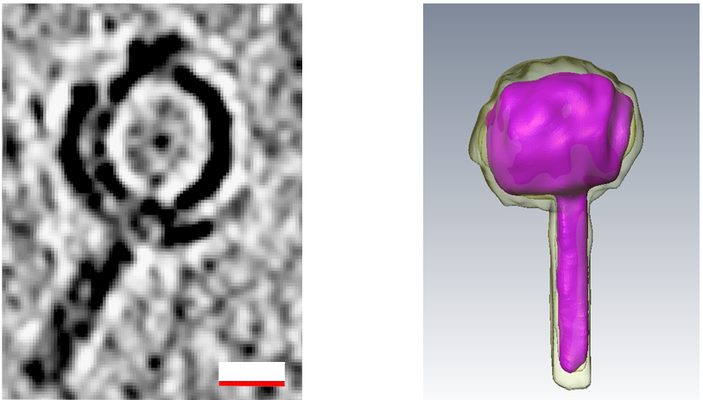 emd_3548.png emd_3548.png | 164 KB | ||
| Others | emd_3548_additional.map.gz | 9.7 MB | ||
| Archive directory |  http://ftp.pdbj.org/pub/emdb/structures/EMD-3548ftp://ftp.pdbj.org/pub/emdb/structures/EMD-3548 http://ftp.pdbj.org/pub/emdb/structures/EMD-3548ftp://ftp.pdbj.org/pub/emdb/structures/EMD-3548 | HTTPS FTP |
-Related structure data

-Links
| EMDB pages | EMDB (EBI/PDBe) / EMDataResource |
|---|
-Map
| File | Download / File: emd_3548.map.gz / Format: CCP4 / Size: 10.5 MB / Type: IMAGE STORED AS FLOATING POINT NUMBER (4 BYTES) | ||||||||||||||||||||||||||||||||||||||||||||||||||||||||||||
|---|---|---|---|---|---|---|---|---|---|---|---|---|---|---|---|---|---|---|---|---|---|---|---|---|---|---|---|---|---|---|---|---|---|---|---|---|---|---|---|---|---|---|---|---|---|---|---|---|---|---|---|---|---|---|---|---|---|---|---|---|---|
| Annotation | Please open in Imagej or Amira | ||||||||||||||||||||||||||||||||||||||||||||||||||||||||||||
| Projections & slices | Image control
Images are generated by Spider. | ||||||||||||||||||||||||||||||||||||||||||||||||||||||||||||
| Voxel size | X=Y=Z: 8.8 Å | ||||||||||||||||||||||||||||||||||||||||||||||||||||||||||||
| Density |
| ||||||||||||||||||||||||||||||||||||||||||||||||||||||||||||
| Symmetry | Space group: 1 | ||||||||||||||||||||||||||||||||||||||||||||||||||||||||||||
| Details | EMDB XML:
CCP4 map header:
| ||||||||||||||||||||||||||||||||||||||||||||||||||||||||||||
 Z (Sec.)
 Y (Row.)
 X (Col.)


-Supplemental data
-Additional map: Denoised volume using TOMOAND
| File | emd_3548_additional.map | ||||||||||||
|---|---|---|---|---|---|---|---|---|---|---|---|---|---|
| Annotation | Denoised volume using TOMOAND | ||||||||||||
| Projections & Slices |
| ||||||||||||
| Density Histograms |


- Sample components
-Entire : Enterobacteria phage PRD1
| Entire | Name: Enterobacteria phage PRD1 (virus) |
|---|---|
| Components |
|
-Supramolecule #1: Enterobacteria phage PRD1
| Supramolecule | Name: Enterobacteria phage PRD1 / type: virus / ID: 1 / Parent: 0 / NCBI-ID: 10658 / Sci species name: Enterobacteria phage PRD1 / Virus type: VIRION / Virus isolate: SPECIES / Virus enveloped: No / Virus empty: No |
|---|---|
| Host (natural) | Organism:  Salmonella enterica subsp. enterica serovar Typhimurium str. LT2 (bacteria) Salmonella enterica subsp. enterica serovar Typhimurium str. LT2 (bacteria) |
| Molecular weight | Experimental: 66 MDa |
| Virus shell | Shell ID: 1 / Name: PRD1 / Diameter: 650.0 Å / T number (triangulation number): 25 |
-Experimental details
-Structure determination
| Method | cryo EM |
|---|---|
 Processing Processing | electron tomography |
| Aggregation state | particle |
-Sample preparation
| Concentration | 0.6 mg/mL | ||||||
|---|---|---|---|---|---|---|---|
| Buffer | pH: 7.2 / Component:
| ||||||
| Grid | Model: QUANTIFOIL R 2/1 (or R 3.5/1) / Material: COPPER / Mesh: 200 | ||||||
| Vitrification | Cryogen name: ETHANE / Chamber humidity: 95 % / Instrument: FEI VITROBOT MARK III / Details: Blot for 4 seconds before plunging. | ||||||
| Details | 20 mM Phosphate Buffer 1 mM MgCl2 | ||||||
| Sectioning | Other: NO SECTIONING | ||||||
| Fiducial marker | Manufacturer: Aurion BSA gold tracer / Diameter: 10 nm |
- Electron microscopy
| Microscope | JEOL 2200FSC |
|---|---|
| Specialist optics | Energy filter - Name: In-column Omega Filter |
| Image recording | Film or detector model: GATAN ULTRASCAN 4000 (4k x 4k) / Average electron dose: 1.3 e/Å2 |
| Electron beam | Acceleration voltage: 200 kV / Electron source:  FIELD EMISSION GUN FIELD EMISSION GUN |
| Electron optics | Calibrated defocus min: 5.0 µm / Illumination mode: FLOOD BEAM / Imaging mode: BRIGHT FIELD / Cs: 2.0 mm / Nominal defocus max: 8.0 µm / Nominal magnification: 25000 |
| Sample stage | Cooling holder cryogen: NITROGEN |
-Image processing
| Final reconstruction | Algorithm: BACK PROJECTION / Software - Name: IMOD / Number images used: 85 |
|---|
