 Movie
Movie Controller


+ Open data

- Basic information
| Entry | 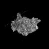 | |||||||||
|---|---|---|---|---|---|---|---|---|---|---|
| Title | Yeast 40S ribosomal subunit | |||||||||
 Map data Map data | Cryo-EM map of yeast 40S ribosomal subunit | |||||||||
 Sample Sample |
| |||||||||
 Keywords Keywords | Yeast small ribosomal subunit 40S / RIBOSOME | |||||||||
| Biological species |  Kluyveromyces lactis NRRL Y-1140 (yeast) Kluyveromyces lactis NRRL Y-1140 (yeast) | |||||||||
| Method | single particle reconstruction / cryo EM / Resolution: 4.0 Å | |||||||||
 Authors Authors | Datey A / Khaja FT / Hussain T | |||||||||
| Funding support |  India, 1 items India, 1 items
| |||||||||
 Citation Citation | Journal: Biochemistry / Year: 2025 Title: Yeast Eukaryotic Initiation Factor 4B Remodels the MRNA Entry Site on the Small Ribosomal Subunit. Authors: Ayushi Datey / Prafful Sharma / Faisal Tarique Khaja / Huma Rahil / Tanweer Hussain / Abstract: Eukaryotic Initiation Factor 4 (eIF4) is a group of factors that activates mRNA for translation and recruit 43S preinitiation complex (PIC) to the mRNA 5' end, forming the 48S PIC. The eIF4 factors ...Eukaryotic Initiation Factor 4 (eIF4) is a group of factors that activates mRNA for translation and recruit 43S preinitiation complex (PIC) to the mRNA 5' end, forming the 48S PIC. The eIF4 factors include mRNA 5' cap-binding protein eIF4E, ATP-dependent RNA helicase eIF4A, and scaffold protein eIF4G, which anchors eIF4A and eIF4E. Another eIF4 factor, eIF4B, stimulates the RNA helicase activity of eIF4A and facilitates mRNA recruitment. However, the mechanisms by which eIF4B binds the 40S ribosomal subunit and promotes mRNA recruitment remain poorly understood. Using cryo-Eletron Microscopy (cryo-EM), we obtained a map of the yeast 40S ribosomal subunit in a complex with eIF4B (40S-eIF4B complex). An extra density, tentatively assigned to yeast eIF4B, was observed near the mRNA entry channel of the 40S, contacting ribosomal proteins uS10, uS3, and eS10 as well as rRNA helix h16. Predictive modeling of the 40S-eIF4B complex suggests that the N-terminal domain of eIF4B binds near the mRNA entry channel, overlapping with the extra density observed in the 40S-eIF4B map. The partially open conformation of 40S in the 40S-eIF4B map is incompatible with eIF3j binding observed in the 48S PIC. Additionally, the extra density at the mRNA entry channel poses steric hindrance for eIF3g binding in the 48S PIC. Thus, structural insights suggest that eIF4B facilitates the release of eIF3j and the relocation of the eIF3b-g-i module during mRNA recruitment, thereby advancing our understanding of eIF4B's role in translation initiation. | |||||||||
| History |
|
- Structure visualization
| Supplemental images |
|---|

- Downloads & links
-EMDB archive
| Map data | emd_35233.map.gz | 167 MB |  EMDB map data format EMDB map data format | |
|---|---|---|---|---|
| Header (meta data) | emd-35233-v30.xmlemd-35233.xml | 19.9 KB 19.9 KB | Display Display | EMDB header |
| FSC (resolution estimation) | emd_35233_fsc.xml | 16.5 KB | Display | FSC data file |
| Images |  emd_35233.png emd_35233.png | 57.4 KB | ||
| Filedesc metadata | emd-35233.cif.gz | 5 KB | ||
| Others | emd_35233_additional_1.map.gzemd_35233_half_map_1.map.gzemd_35233_half_map_2.map.gz | 139.5 MB 140.9 MB 140.9 MB | ||
| Archive directory |  http://ftp.pdbj.org/pub/emdb/structures/EMD-35233ftp://ftp.pdbj.org/pub/emdb/structures/EMD-35233 http://ftp.pdbj.org/pub/emdb/structures/EMD-35233ftp://ftp.pdbj.org/pub/emdb/structures/EMD-35233 | HTTPS FTP |
-Related structure data

-Links
| EMDB pages | EMDB (EBI/PDBe) / EMDataResource |
|---|
-Map
| File | Download / File: emd_35233.map.gz / Format: CCP4 / Size: 178 MB / Type: IMAGE STORED AS FLOATING POINT NUMBER (4 BYTES) | ||||||||||||||||||||||||||||||||||||
|---|---|---|---|---|---|---|---|---|---|---|---|---|---|---|---|---|---|---|---|---|---|---|---|---|---|---|---|---|---|---|---|---|---|---|---|---|---|
| Annotation | Cryo-EM map of yeast 40S ribosomal subunit | ||||||||||||||||||||||||||||||||||||
| Projections & slices | Image control
Images are generated by Spider. | ||||||||||||||||||||||||||||||||||||
| Voxel size | X=Y=Z: 1.17 Å | ||||||||||||||||||||||||||||||||||||
| Density |
| ||||||||||||||||||||||||||||||||||||
| Symmetry | Space group: 1 | ||||||||||||||||||||||||||||||||||||
| Details | EMDB XML:
|
 Z (Sec.)
 Y (Row.)
 X (Col.)


-Supplemental data
-Additional map: Auto-refined map of yeast 40S ribosomal subunit.
| File | emd_35233_additional_1.map | ||||||||||||
|---|---|---|---|---|---|---|---|---|---|---|---|---|---|
| Annotation | Auto-refined map of yeast 40S ribosomal subunit. | ||||||||||||
| Projections & Slices |
| ||||||||||||
| Density Histograms |


-Half map: Half map 1 of yeast 40S ribosomal subunit
| File | emd_35233_half_map_1.map | ||||||||||||
|---|---|---|---|---|---|---|---|---|---|---|---|---|---|
| Annotation | Half map 1 of yeast 40S ribosomal subunit | ||||||||||||
| Projections & Slices |
| ||||||||||||
| Density Histograms |


-Half map: Half map 2 of yeast 40S ribosomal subunit
| File | emd_35233_half_map_2.map | ||||||||||||
|---|---|---|---|---|---|---|---|---|---|---|---|---|---|
| Annotation | Half map 2 of yeast 40S ribosomal subunit | ||||||||||||
| Projections & Slices |
| ||||||||||||
| Density Histograms |


- Sample components
-Entire : 40S subunit of yeast
| Entire | Name: 40S subunit of yeast |
|---|---|
| Components |
|
-Supramolecule #1: 40S subunit of yeast
| Supramolecule | Name: 40S subunit of yeast / type: complex / ID: 1 / Parent: 0 Details: This 40S cryo-EM map was obtained during masked and unmasked 3D classification of the cryo-EM dataset of the 40S-eIF4B complex |
|---|---|
| Source (natural) | Organism: Kluyveromyces lactis NRRL Y-1140 (yeast) / Strain: NRRL Y-1140 |
-Experimental details
-Structure determination
| Method | cryo EM |
|---|---|
 Processing Processing | single particle reconstruction |
| Aggregation state | particle |
-Sample preparation
| Buffer | pH: 7.4 Component:
Details: 20 mM HEPES pH7.4, 100 mM potassium acetate pH7.6, 2.5 mM Magnesium acetate, 2 mM DTT | |||||||||||||||
|---|---|---|---|---|---|---|---|---|---|---|---|---|---|---|---|---|
| Grid | Model: Quantifoil R1.2/1.3 / Material: COPPER / Mesh: 300 / Support film - Material: CARBON / Support film - topology: CONTINUOUS / Pretreatment - Type: GLOW DISCHARGE / Pretreatment - Time: 90 sec. / Pretreatment - Atmosphere: AIR / Pretreatment - Pressure: 100.0 kPa | |||||||||||||||
| Vitrification | Cryogen name: ETHANE / Chamber humidity: 100 % / Chamber temperature: 295 K / Instrument: FEI VITROBOT MARK IV Details: Vitrification was performed in nitrogen atmosphere. | |||||||||||||||
| Details | This class of empty or only 40S was a part of the reaction mixture containing 40S and the Saccharomyces cerevisiae eukaryotic initiation factor 4B present in a 1:10 molar ratio |
- Electron microscopy
| Microscope | FEI TALOS ARCTICA |
|---|---|
| Temperature | Min: 77.0 K / Max: 98.0 K |
| Details | Preliminary grid screening was done manually using the Latitude-S |
| Image recording | Film or detector model: GATAN K2 SUMMIT (4k x 4k) / Detector mode: COUNTING / Digitization - Frames/image: 4-16 / Number grids imaged: 6 / Number real images: 7975 / Average exposure time: 8.0 sec. / Average electron dose: 50.0 e/Å2 Details: Images were collected in movie-mode at 20 frames per 8 seconds |
| Electron beam | Acceleration voltage: 200 kV / Electron source:  FIELD EMISSION GUN FIELD EMISSION GUN |
| Electron optics | C2 aperture diameter: 70.0 µm / Calibrated defocus max: 3.0 µm / Calibrated defocus min: 1.5 µm / Calibrated magnification: 42000 / Illumination mode: OTHER / Imaging mode: DIFFRACTION / Cs: 2.7 mm / Nominal defocus max: 3.0 µm / Nominal defocus min: 1.5 µm / Nominal magnification: 42000 |
| Sample stage | Cooling holder cryogen: NITROGEN |
| Experimental equipment |  Model: Talos Arctica / Image courtesy: FEI Company |
+Image processing


-Atomic model buiding 1
| Refinement | Protocol: RIGID BODY FIT / Target criteria: Cross-correlation coefficient |
|---|
