 Movie
Movie Controller


+ Open data

- Basic information
| Entry | 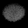 | |||||||||||||||
|---|---|---|---|---|---|---|---|---|---|---|---|---|---|---|---|---|
| Title | The asymmetric unit of P22 empty capsid | |||||||||||||||
 Map data Map data | ||||||||||||||||
 Sample Sample |
| |||||||||||||||
 Keywords Keywords | Complex / VIRUS / VIRAL PROTEIN | |||||||||||||||
| Function / homology | Major capsid protein Gp5 / P22 coat protein - gene protein 5 / viral procapsid / viral procapsid maturation / T=7 icosahedral viral capsid / viral capsid / identical protein binding / Major capsid protein Function and homology information Function and homology information | |||||||||||||||
| Biological species |  Salmonella phage P22 (virus) Salmonella phage P22 (virus) | |||||||||||||||
| Method | single particle reconstruction / cryo EM / Resolution: 2.8 Å | |||||||||||||||
 Authors Authors | Xiao H / Liu HR / Cheng LP | |||||||||||||||
| Funding support |  China, 4 items China, 4 items
| |||||||||||||||
 Citation Citation | Journal: Viruses / Year: 2023 Title: Assembly and Capsid Expansion Mechanism of Bacteriophage P22 Revealed by High-Resolution Cryo-EM Structures. Authors: Hao Xiao / Junquan Zhou / Fan Yang / Zheng Liu / Jingdong Song / Wenyuan Chen / Hongrong Liu / Lingpeng Cheng / Abstract: The formation of many double-stranded DNA viruses, such as herpesviruses and bacteriophages, begins with the scaffolding-protein-mediated assembly of the procapsid. Subsequently, the procapsid ...The formation of many double-stranded DNA viruses, such as herpesviruses and bacteriophages, begins with the scaffolding-protein-mediated assembly of the procapsid. Subsequently, the procapsid undergoes extensive structural rearrangement and expansion to become the mature capsid. Bacteriophage P22 is an established model system used to study virus maturation. Here, we report the cryo-electron microscopy structures of procapsid, empty procapsid, empty mature capsid, and mature capsid of phage P22 at resolutions of 2.6 Å, 3.9 Å, 2.8 Å, and 3.0 Å, respectively. The structure of the procapsid allowed us to build an accurate model of the coat protein gp5 and the C-terminal region of the scaffolding protein gp8. In addition, interactions among the gp5 subunits responsible for procapsid assembly and stabilization were identified. Two C-terminal α-helices of gp8 were observed to interact with the coat protein in the procapsid. The amino acid interactions between gp5 and gp8 in the procapsid were consistent with the results of previous biochemical studies involving mutant proteins. Our structures reveal hydrogen bonds and salt bridges between the gp5 subunits in the procapsid and the conformational changes of the gp5 domains involved in the closure of the local sixfold opening and a thinner capsid shell during capsid maturation. | |||||||||||||||
| History |
|
- Structure visualization
| Supplemental images |
|---|

- Downloads & links
-EMDB archive
| Map data | emd_35121.map.gz | 165.2 MB | EMDB map data format | |
|---|---|---|---|---|
| Header (meta data) | emd-35121-v30.xmlemd-35121.xml | 16.8 KB 16.8 KB | Display Display | EMDB header |
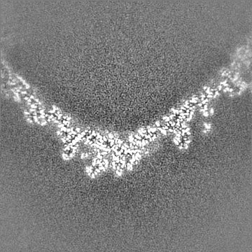
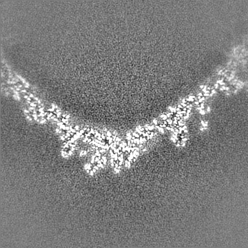
| Images |  emd_35121.png emd_35121.png | 195.4 KB | ||
| Filedesc metadata | emd-35121.cif.gz | 5.7 KB | ||
| Others | emd_35121_half_map_1.map.gzemd_35121_half_map_2.map.gz | 164.5 MB 164.5 MB | ||
| Archive directory |  http://ftp.pdbj.org/pub/emdb/structures/EMD-35121ftp://ftp.pdbj.org/pub/emdb/structures/EMD-35121 http://ftp.pdbj.org/pub/emdb/structures/EMD-35121ftp://ftp.pdbj.org/pub/emdb/structures/EMD-35121 | HTTPS FTP |
-Validation report
| Summary document | emd_35121_validation.pdf.gz | 1.2 MB | Display | EMDB validaton report |
|---|---|---|---|---|
| Full document | emd_35121_full_validation.pdf.gz | 1.2 MB | Display | |
| Data in XML | emd_35121_validation.xml.gz | 15.3 KB | Display | |
| Data in CIF | emd_35121_validation.cif.gz | 18.3 KB | Display | |
| Arichive directory | https://ftp.pdbj.org/pub/emdb/validation_reports/EMD-35121ftp://ftp.pdbj.org/pub/emdb/validation_reports/EMD-35121 | HTTPS FTP |
-Related structure data
| Related structure data |  8i1tMC  8i1vC M: atomic model generated by this map C: citing same article ( |
|---|---|
| Similar structure data |


-Links
| EMDB pages | EMDB (EBI/PDBe) / EMDataResource |
|---|---|
| Related items in Molecule of the Month |

-Map
| File | Download / File: emd_35121.map.gz / Format: CCP4 / Size: 178 MB / Type: IMAGE STORED AS FLOATING POINT NUMBER (4 BYTES) | ||||||||||||||||||||||||||||||||||||
|---|---|---|---|---|---|---|---|---|---|---|---|---|---|---|---|---|---|---|---|---|---|---|---|---|---|---|---|---|---|---|---|---|---|---|---|---|---|
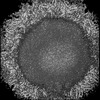
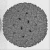
| Projections & slices | Image control
Images are generated by Spider. | ||||||||||||||||||||||||||||||||||||
| Voxel size | X=Y=Z: 1.06 Å | ||||||||||||||||||||||||||||||||||||
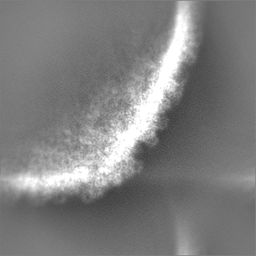
| Density |
| ||||||||||||||||||||||||||||||||||||
| Symmetry | Space group: 1 | ||||||||||||||||||||||||||||||||||||
| Details | EMDB XML:
|
 Z (Sec.)
 Y (Row.)
 X (Col.)


-Supplemental data
-Half map: #2
| File | emd_35121_half_map_1.map | ||||||||||||
|---|---|---|---|---|---|---|---|---|---|---|---|---|---|
| Projections & Slices |
| ||||||||||||
| Density Histograms |


-Half map: #1
| File | emd_35121_half_map_2.map | ||||||||||||
|---|---|---|---|---|---|---|---|---|---|---|---|---|---|
| Projections & Slices |
| ||||||||||||
| Density Histograms |


- Sample components
-Entire : Salmonella phage P22
| Entire | Name: Salmonella phage P22 (virus) |
|---|---|
| Components |
|
-Supramolecule #1: Salmonella phage P22
| Supramolecule | Name: Salmonella phage P22 / type: virus / ID: 1 / Parent: 0 / Macromolecule list: all / NCBI-ID: 2908168 / Sci species name: Salmonella phage P22 / Virus type: VIRION / Virus isolate: SPECIES / Virus enveloped: No / Virus empty: Yes |
|---|
-Macromolecule #1: Major capsid protein
| Macromolecule | Name: Major capsid protein / type: protein_or_peptide / ID: 1 / Number of copies: 7 / Enantiomer: LEVO |
|---|---|
| Source (natural) | Organism: Salmonella phage P22 (virus) |
| Molecular weight | Theoretical: 46.795613 KDa |
| Sequence | String: MALNEGQIVT LAVDEIIETI SAITPMAQKA KKYTPPAASM QRSSNTIWMP VEQESPTQEG WDLTDKATGL LELNVAVNMG EPDNDFFQL RADDLRDETA YRRRIQSAAR KLANNVELKV ANMAAEMGSL VITSPDAIGT NTADAWNFVA DAEEIMFSRE L NRDMGTSY ...String: MALNEGQIVT LAVDEIIETI SAITPMAQKA KKYTPPAASM QRSSNTIWMP VEQESPTQEG WDLTDKATGL LELNVAVNMG EPDNDFFQL RADDLRDETA YRRRIQSAAR KLANNVELKV ANMAAEMGSL VITSPDAIGT NTADAWNFVA DAEEIMFSRE L NRDMGTSY FFNPQDYKKA GYDLTKRDIF GRIPEEAYRD GTIQRQVAGF DDVLRSPKLP VLTKSTATGI TVSGAQSFKP VA WQLDNDG NKVNVDNRFA TVTLSATTGM KRGDKISFAG VKFLGQMAKN VLAQDATFSV VRVVDGTHVE ITPKPVALDD VSL SPEQRA YANVNTSLAD AMAVNILNVK DARTNVFWAD DAIRIVSQPI PANHELFAGM KTTSFSIPDV GLNGIFATQG DIST LSGLC RIALWYGVNA TRPEAIGVGL PGQTA UniProtKB: Major capsid protein |
-Experimental details
-Structure determination
| Method | cryo EM |
|---|---|
 Processing Processing | single particle reconstruction |
| Aggregation state | particle |
-Sample preparation
| Buffer | pH: 7.4 |
|---|---|
| Vitrification | Cryogen name: ETHANE |
- Electron microscopy
| Microscope | FEI TITAN KRIOS |
|---|---|
| Image recording | Film or detector model: GATAN K3 (6k x 4k) / Average electron dose: 35.0 e/Å2 |
| Electron beam | Acceleration voltage: 300 kV / Electron source:  FIELD EMISSION GUN FIELD EMISSION GUN |
| Electron optics | Illumination mode: FLOOD BEAM / Imaging mode: BRIGHT FIELD / Nominal defocus max: 2.4 µm / Nominal defocus min: 1.6 µm |
| Experimental equipment |  Model: Titan Krios / Image courtesy: FEI Company |
