National Natural Science Foundation of China (NSFC)
12034006
China
National Natural Science Foundation of China (NSFC)
31971122
China
National Natural Science Foundation of China (NSFC)
32071209
China
National Natural Science Foundation of China (NSFC)
32200994
China
Citation
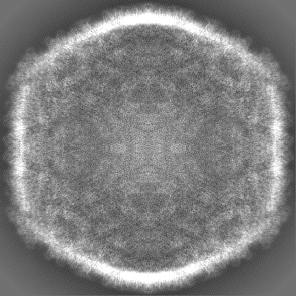
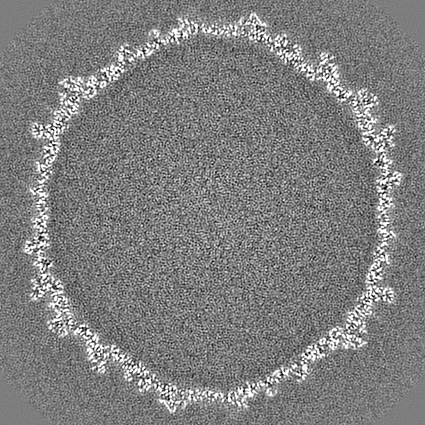
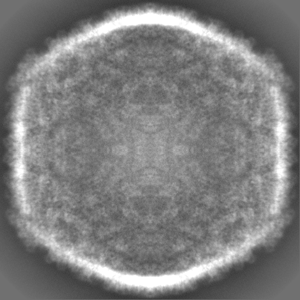
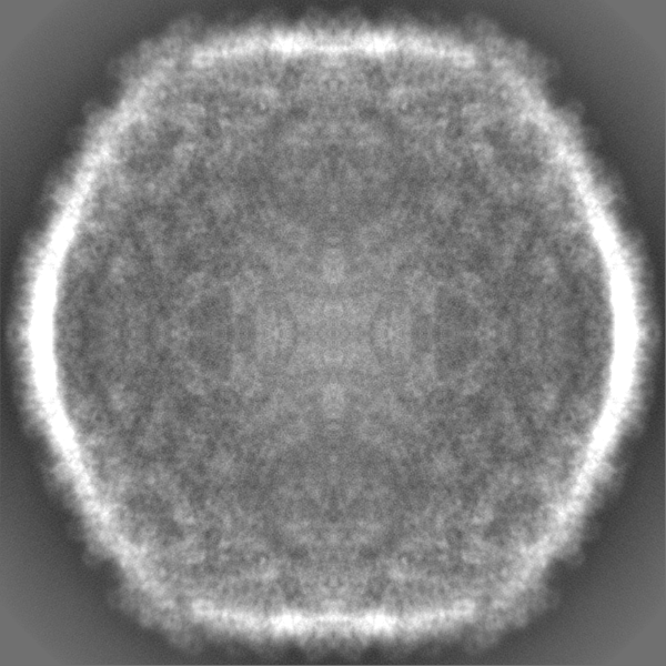
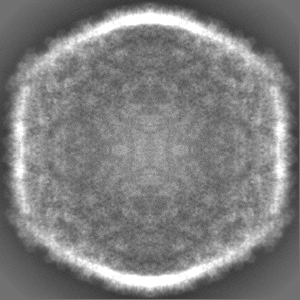
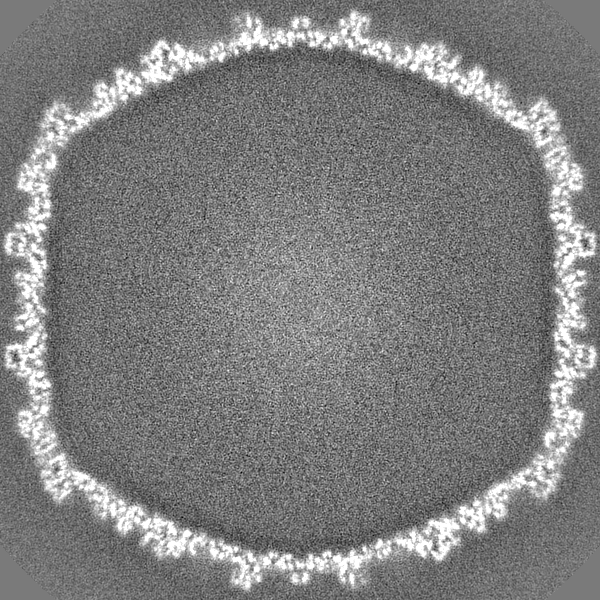
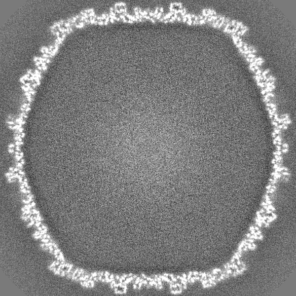
Journal: Viruses / Year: 2023 Title: Assembly and Capsid Expansion Mechanism of Bacteriophage P22 Revealed by High-Resolution Cryo-EM Structures. Authors: Hao Xiao / Junquan Zhou / Fan Yang / Zheng Liu / Jingdong Song / Wenyuan Chen / Hongrong Liu / Lingpeng Cheng / Abstract: The formation of many double-stranded DNA viruses, such as herpesviruses and bacteriophages, begins with the scaffolding-protein-mediated assembly of the procapsid. Subsequently, the procapsid ...The formation of many double-stranded DNA viruses, such as herpesviruses and bacteriophages, begins with the scaffolding-protein-mediated assembly of the procapsid. Subsequently, the procapsid undergoes extensive structural rearrangement and expansion to become the mature capsid. Bacteriophage P22 is an established model system used to study virus maturation. Here, we report the cryo-electron microscopy structures of procapsid, empty procapsid, empty mature capsid, and mature capsid of phage P22 at resolutions of 2.6 Å, 3.9 Å, 2.8 Å, and 3.0 Å, respectively. The structure of the procapsid allowed us to build an accurate model of the coat protein gp5 and the C-terminal region of the scaffolding protein gp8. In addition, interactions among the gp5 subunits responsible for procapsid assembly and stabilization were identified. Two C-terminal α-helices of gp8 were observed to interact with the coat protein in the procapsid. The amino acid interactions between gp5 and gp8 in the procapsid were consistent with the results of previous biochemical studies involving mutant proteins. Our structures reveal hydrogen bonds and salt bridges between the gp5 subunits in the procapsid and the conformational changes of the gp5 domains involved in the closure of the local sixfold opening and a thinner capsid shell during capsid maturation.
In the structure databanks used in Yorodumi, some data are registered as the other names, "COVID-19 virus" and "2019-nCoV". Here are the details of the virus and the list of structure data.
Jan 31, 2019. EMDB accession codes are about to change! (news from PDBe EMDB page)
EMDB accession codes are about to change! (news from PDBe EMDB page)
The allocation of 4 digits for EMDB accession codes will soon come to an end. Whilst these codes will remain in use, new EMDB accession codes will include an additional digit and will expand incrementally as the available range of codes is exhausted. The current 4-digit format prefixed with “EMD-” (i.e. EMD-XXXX) will advance to a 5-digit format (i.e. EMD-XXXXX), and so on. It is currently estimated that the 4-digit codes will be depleted around Spring 2019, at which point the 5-digit format will come into force.
The EM Navigator/Yorodumi systems omit the EMD- prefix.
Related info.:Q: What is EMD? / ID/Accession-code notation in Yorodumi/EM Navigator
Yorodumi is a browser for structure data from EMDB, PDB, SASBDB, etc.
This page is also the successor to EM Navigator detail page, and also detail information page/front-end page for Omokage search.
The word "yorodu" (or yorozu) is an old Japanese word meaning "ten thousand". "mi" (miru) is to see.
Related info.:EMDB / PDB / SASBDB / Comparison of 3 databanks / Yorodumi Search / Aug 31, 2016. New EM Navigator & Yorodumi / Yorodumi Papers / Jmol/JSmol / Function and homology information / Changes in new EM Navigator and Yorodumi
 Movie
Movie Controller


 Open data
 Basic information
 Map data
 Sample
 Keywords
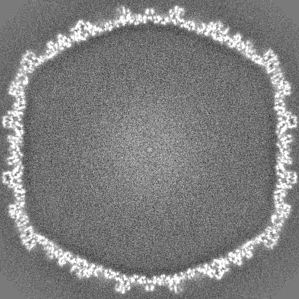
 Salmonella phage P22 (virus)
 Authors
 China, 4 items
  Citation
 Structure visualization
 Downloads & links
 EMDB map data format
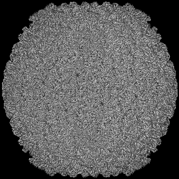
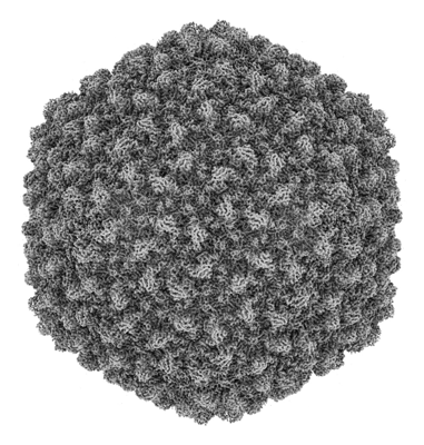 emd_35120.png
 http://ftp.pdbj.org/pub/emdb/structures/EMD-35120


 Z (Sec.)
 Y (Row.)
 X (Col.)


 Sample components
 Processing
 Electron microscopy
 FIELD EMISSION GUN
