ムービー
ムービー コントローラー


+ データを開く

- 基本情報
基本情報
| 登録情報 |  | |||||||||
|---|---|---|---|---|---|---|---|---|---|---|
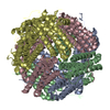
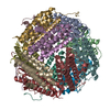
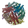
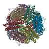
| タイトル | Cryo-EM structure of MsDps2 from Mycobacterium smegmatis | |||||||||
 マップデータ マップデータ | ||||||||||
 試料 試料 |
| |||||||||
 キーワード キーワード | MsDps2 / cryo-EM / DNA BINDING PROTEIN | |||||||||
| 機能・相同性 |  機能・相同性情報 機能・相同性情報 | |||||||||
| 生物種 |  Mycolicibacterium smegmatis MC2 155 (バクテリア) Mycolicibacterium smegmatis MC2 155 (バクテリア) | |||||||||
| 手法 | 単粒子再構成法 / クライオ電子顕微鏡法 / 解像度: 2.9 Å | |||||||||
 データ登録者 データ登録者 | Garg P / Dutta S | |||||||||
| 資金援助 |  インド, 2件 インド, 2件
| |||||||||
 引用 引用 | ジャーナル: J Mol Biol / 年: 2024 タイトル: Cryo-EM Reveals the Mechanism of DNA Compaction by Mycobacterium smegmatis Dps2. 著者: Priyanka Garg / Thejas Satheesh / Mahipal Ganji / Somnath Dutta / 要旨: DNA binding protein from starved cells (Dps) is a miniature ferritin complex, which plays a vital role in protecting bacterial DNA during starvation to maintain the integrity of bacteria under ...DNA binding protein from starved cells (Dps) is a miniature ferritin complex, which plays a vital role in protecting bacterial DNA during starvation to maintain the integrity of bacteria under hostile conditions. Several approaches, including cryo-electron tomography, have been previously implemented by other research groups to decipher the structure of the Dps protein bound to DNA. However, none of the structures of the Dps-DNA complex was resolved to high resolution to identify the DNA binding residues. Like other bacteria, Mycobacterium smegmatis also expresses Dps2 (called MsDps2), which binds DNA to protect it under oxidative stress conditions. In this study, we implemented various biochemical and biophysical studies to characterize the DNA protein interactions of Dps2 protein from Mycobacterium smegmatis. We employed single-particle cryo-EM-based structural analysis of MsDps2-DNA complexes and identified that the region close to the N-terminus confers the DNA binding property. Based on cryo-EM data, we also pinpointed several arginine residues, proximal to the DNA binding region, responsible for DNA binding. We also performed mutations of these residues, which dramatically reduced the MsDps2-DNA interaction. In addition, we proposed a model that elucidates the mechanism of DNA compaction, which adapts a lattice-like structure. We performed single-molecule imaging of MsDps2-DNA interactions that corroborate well with our structural studies. Taken together, our results delineate the specific MsDps2 residues that play an important role in DNA binding and compaction, providing new insights into Mycobacterial DNA compaction mechanisms under stress conditions. | |||||||||
| 履歴 |
|
- 構造の表示
構造の表示
- ダウンロードとリンク
-EMDBアーカイブ
| マップデータ | emd_35070.map.gz | 126.3 MB | EMDBマップデータ形式 | |
|---|---|---|---|---|
| ヘッダ (付随情報) | emd-35070-v30.xmlemd-35070.xml | 16.5 KB 16.5 KB | 表示 表示 | EMDBヘッダ |
| FSC (解像度算出) | emd_35070_fsc.xml | 10.8 KB | 表示 | FSCデータファイル |
| 画像 |  emd_35070.png emd_35070.png | 140.4 KB | ||
| Filedesc metadata | emd-35070.cif.gz | 5.7 KB | ||
| その他 | emd_35070_half_map_1.map.gzemd_35070_half_map_2.map.gz | 124.6 MB 124.6 MB | ||
| アーカイブディレクトリ |  http://ftp.pdbj.org/pub/emdb/structures/EMD-35070ftp://ftp.pdbj.org/pub/emdb/structures/EMD-35070 http://ftp.pdbj.org/pub/emdb/structures/EMD-35070ftp://ftp.pdbj.org/pub/emdb/structures/EMD-35070 | HTTPS FTP |
-検証レポート
| 文書・要旨 | emd_35070_validation.pdf.gz | 1 MB | 表示 | EMDB検証レポート |
|---|---|---|---|---|
| 文書・詳細版 | emd_35070_full_validation.pdf.gz | 1 MB | 表示 | |
| XML形式データ | emd_35070_validation.xml.gz | 19.6 KB | 表示 | |
| CIF形式データ | emd_35070_validation.cif.gz | 25.2 KB | 表示 | |
| アーカイブディレクトリ | https://ftp.pdbj.org/pub/emdb/validation_reports/EMD-35070ftp://ftp.pdbj.org/pub/emdb/validation_reports/EMD-35070 | HTTPS FTP |
-関連構造データ


-リンク
| EMDBのページ | EMDB (EBI/PDBe) / EMDataResource |
|---|---|
| 「今月の分子」の関連する項目 |


-マップ
| ファイル | ダウンロード / ファイル: emd_35070.map.gz / 形式: CCP4 / 大きさ: 134.6 MB / タイプ: IMAGE STORED AS FLOATING POINT NUMBER (4 BYTES) | ||||||||||||||||||||
|---|---|---|---|---|---|---|---|---|---|---|---|---|---|---|---|---|---|---|---|---|---|
| ボクセルのサイズ | X=Y=Z: 0.74 Å | ||||||||||||||||||||
| 密度 |
| ||||||||||||||||||||
| 対称性 | 空間群: 1 | ||||||||||||||||||||
| 詳細 | EMDB XML:
|
-添付データ
- 試料の構成要素
-全体 : Cryo-EM structure of MsDps2 from Mycobacterium Smegmatis
| 全体 | 名称: Cryo-EM structure of MsDps2 from Mycobacterium Smegmatis |
|---|---|
| 要素 |
|
-超分子 #1: Cryo-EM structure of MsDps2 from Mycobacterium Smegmatis
| 超分子 | 名称: Cryo-EM structure of MsDps2 from Mycobacterium Smegmatis タイプ: complex / ID: 1 / 親要素: 0 / 含まれる分子: all |
|---|---|
| 由来(天然) | 生物種: Mycolicibacterium smegmatis MC2 155 (バクテリア) |
-分子 #1: Putative starvation-induced DNA protecting protein/Ferritin and Dps
| 分子 | 名称: Putative starvation-induced DNA protecting protein/Ferritin and Dps タイプ: protein_or_peptide / ID: 1 / コピー数: 12 / 光学異性体: LEVO |
|---|---|
| 由来(天然) | 生物種: Mycolicibacterium smegmatis MC2 155 (バクテリア) |
| 分子量 | 理論値: 17.854863 KDa |
| 組換発現 | 生物種: |
| 配列 | 文字列: MSARRTESDI QGFHATPEFG GNLQKVLVDL IELSLQGKQA HWNVVGSNFR DLHLQLDELV DFAREGSDTI AERMRALDAV PDGRSDTVA ATTTLPEFPA FERSTADVVD LITTRINATV DTIRRVHDAV DAEDPSTADL LHGLIDGLEK QAWLIRSENR K V UniProtKB: Starvation-inducible DNA-binding protein or fine tangled pili major subunit |
-実験情報
-構造解析
| 手法 | クライオ電子顕微鏡法 |
|---|---|
 解析 解析 | 単粒子再構成法 |
| 試料の集合状態 | particle |
-試料調製
| 緩衝液 | pH: 7.5 |
|---|---|
| 凍結 | 凍結剤: ETHANE |
- 電子顕微鏡法
| 顕微鏡 | FEI TALOS ARCTICA |
|---|---|
| 撮影 | フィルム・検出器のモデル: GATAN K2 SUMMIT (4k x 4k) 検出モード: COUNTING / 平均電子線量: 40.0 e/Å2 |
| 電子線 | 加速電圧: 200 kV / 電子線源:  FIELD EMISSION GUN FIELD EMISSION GUN |
| 電子光学系 | 照射モード: OTHER / 撮影モード: BRIGHT FIELD / 最大 デフォーカス(公称値): 2.25 µm / 最小 デフォーカス(公称値): 0.75 µm |
| 実験機器 |  モデル: Talos Arctica / 画像提供: FEI Company |