ムービー
ムービー コントローラー


+ データを開く

- 基本情報
基本情報
| 登録情報 |  | |||||||||
|---|---|---|---|---|---|---|---|---|---|---|
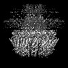
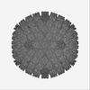
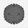
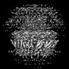
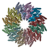
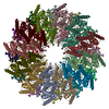
| タイトル | C5 portal vertex in HCMV B-capsid | |||||||||
 マップデータ マップデータ | ||||||||||
 試料 試料 |
| |||||||||
 キーワード キーワード | B-capsid / C5 portal vertex / in-situ structure / VIRUS / VIRAL PROTEIN | |||||||||
| 機能・相同性 |  機能・相同性情報 機能・相同性情報viral genome packaging / T=16 icosahedral viral capsid / viral capsid assembly / viral release from host cell / viral process / chromosome organization / viral capsid / host cell nucleus / structural molecule activity / DNA binding 類似検索 - 分子機能 | |||||||||
| 生物種 |   Human betaherpesvirus 5 (ヘルペスウイルス) Human betaherpesvirus 5 (ヘルペスウイルス) | |||||||||
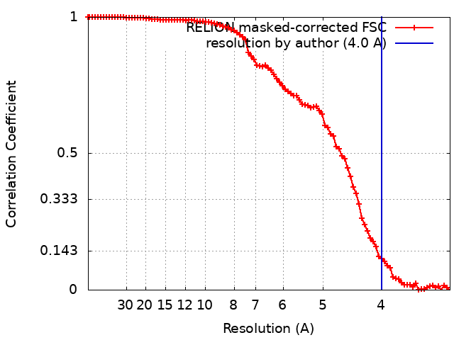
| 手法 | 単粒子再構成法 / クライオ電子顕微鏡法 / 解像度: 4.0 Å | |||||||||
 データ登録者 データ登録者 | Li Z / Yu X | |||||||||
| 資金援助 |  中国, 1件 中国, 1件
| |||||||||
 引用 引用 | ジャーナル: Nat Commun / 年: 2023 タイトル: Cryo-electron microscopy structures of capsids and in situ portals of DNA-devoid capsids of human cytomegalovirus. 著者: Zhihai Li / Jingjing Pang / Rongchao Gao / Qingxia Wang / Maoyan Zhang / Xuekui Yu / 要旨: The portal-scaffold complex is believed to nucleate the assembly of herpesvirus procapsids. During capsid maturation, two events occur: scaffold expulsion and DNA incorporation. The portal-scaffold ...The portal-scaffold complex is believed to nucleate the assembly of herpesvirus procapsids. During capsid maturation, two events occur: scaffold expulsion and DNA incorporation. The portal-scaffold interaction and the conformational changes that occur to the portal during the different stages of capsid formation have yet to be elucidated structurally. Here we present high-resolution structures of the A- and B-capsids and in-situ portals of human cytomegalovirus. We show that scaffolds bind to the hydrophobic cavities formed by the dimerization and Johnson-fold domains of the major capsid proteins. We further show that 12 loop-helix-loop fragments-presumably from the scaffold domain-insert into the hydrophobic pocket of the portal crown domain. The portal also undergoes significant changes both positionally and conformationally as it accompanies DNA packaging. These findings unravel the mechanism by which the portal interacts with the scaffold to nucleate capsid assembly and further our understanding of scaffold expulsion and DNA incorporation. | |||||||||
| 履歴 |
|
- 構造の表示
構造の表示
| 添付画像 |
|---|

- ダウンロードとリンク
-EMDBアーカイブ
| マップデータ | emd_34696.map.gz | 59.8 MB | EMDBマップデータ形式 | |
|---|---|---|---|---|
| ヘッダ (付随情報) | emd-34696-v30.xmlemd-34696.xml | 25.3 KB 25.3 KB | 表示 表示 | EMDBヘッダ |
| FSC (解像度算出) | emd_34696_fsc.xml | 9.1 KB | 表示 | FSCデータファイル |
| 画像 |  emd_34696.png emd_34696.png | 64.6 KB | ||
| Filedesc metadata | emd-34696.cif.gz | 7.9 KB | ||
| その他 | emd_34696_half_map_1.map.gzemd_34696_half_map_2.map.gz | 49.1 MB 49.1 MB | ||
| アーカイブディレクトリ |  http://ftp.pdbj.org/pub/emdb/structures/EMD-34696ftp://ftp.pdbj.org/pub/emdb/structures/EMD-34696 http://ftp.pdbj.org/pub/emdb/structures/EMD-34696ftp://ftp.pdbj.org/pub/emdb/structures/EMD-34696 | HTTPS FTP |
-検証レポート
| 文書・要旨 | emd_34696_validation.pdf.gz | 1.2 MB | 表示 | EMDB検証レポート |
|---|---|---|---|---|
| 文書・詳細版 | emd_34696_full_validation.pdf.gz | 1.2 MB | 表示 | |
| XML形式データ | emd_34696_validation.xml.gz | 16.6 KB | 表示 | |
| CIF形式データ | emd_34696_validation.cif.gz | 21 KB | 表示 | |
| アーカイブディレクトリ | https://ftp.pdbj.org/pub/emdb/validation_reports/EMD-34696ftp://ftp.pdbj.org/pub/emdb/validation_reports/EMD-34696 | HTTPS FTP |
-関連構造データ


-リンク
| EMDBのページ | EMDB (EBI/PDBe) / EMDataResource |
|---|---|
| 「今月の分子」の関連する項目 |

-マップ
| ファイル | ダウンロード / ファイル: emd_34696.map.gz / 形式: CCP4 / 大きさ: 64 MB / タイプ: IMAGE STORED AS FLOATING POINT NUMBER (4 BYTES) | ||||||||||||||||||||||||||||||||||||
|---|---|---|---|---|---|---|---|---|---|---|---|---|---|---|---|---|---|---|---|---|---|---|---|---|---|---|---|---|---|---|---|---|---|---|---|---|---|
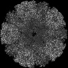
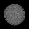
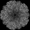
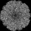
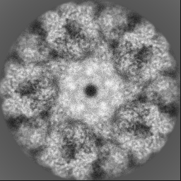
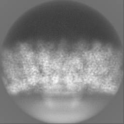
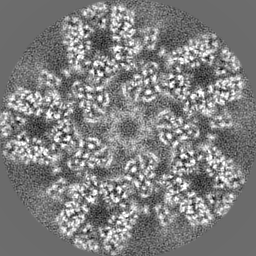
| 投影像・断面図 | 画像のコントロール
画像は Spider により作成 | ||||||||||||||||||||||||||||||||||||
| ボクセルのサイズ | X=Y=Z: 1.625 Å | ||||||||||||||||||||||||||||||||||||
| 密度 |
| ||||||||||||||||||||||||||||||||||||
| 対称性 | 空間群: 1 | ||||||||||||||||||||||||||||||||||||
| 詳細 | EMDB XML:
|
 Z (Sec.)
 Y (Row.)
 X (Col.)


-添付データ
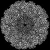
-ハーフマップ: #2
| ファイル | emd_34696_half_map_1.map | ||||||||||||
|---|---|---|---|---|---|---|---|---|---|---|---|---|---|
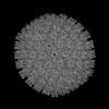
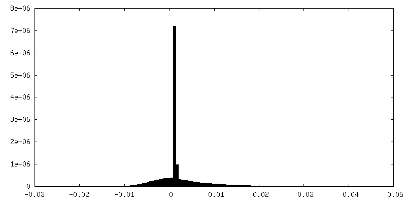
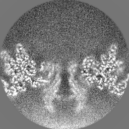
| 投影像・断面図 |
| ||||||||||||
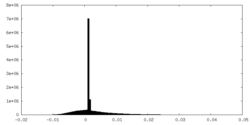
| 密度ヒストグラム |


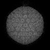
-ハーフマップ: #1
| ファイル | emd_34696_half_map_2.map | ||||||||||||
|---|---|---|---|---|---|---|---|---|---|---|---|---|---|
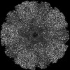
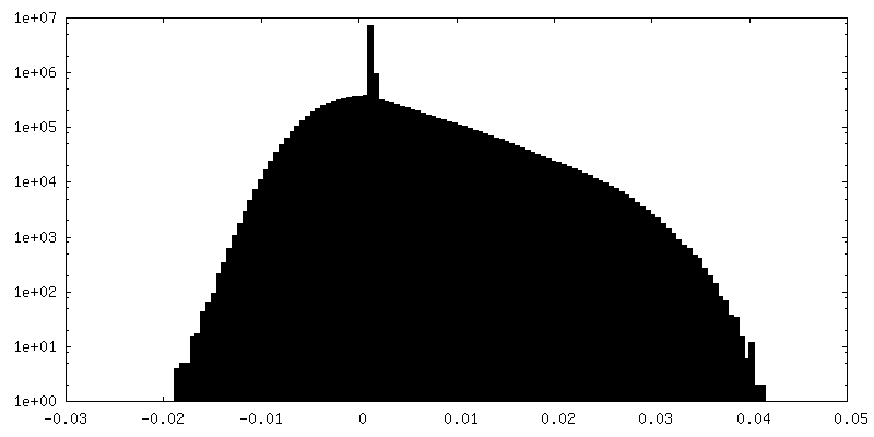
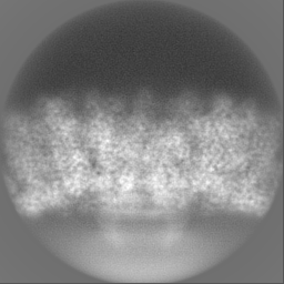
| 投影像・断面図 |
| ||||||||||||
| 密度ヒストグラム |


- 試料の構成要素
+全体 : Human betaherpesvirus 5
+超分子 #1: Human betaherpesvirus 5
+分子 #1: Triplex capsid protein 2
+分子 #2: Triplex capsid protein 1
+分子 #3: Capsid vertex component 1
+分子 #4: Capsid vertex component 2
+分子 #5: Small capsomere-interacting protein
+分子 #6: Major capsid protein
+分子 #7: portal protein
+分子 #8: portal protein
+分子 #9: portal protein
-実験情報
-構造解析
| 手法 | クライオ電子顕微鏡法 |
|---|---|
 解析 解析 | 単粒子再構成法 |
| 試料の集合状態 | particle |
-試料調製
| 緩衝液 | pH: 7.4 |
|---|---|
| 凍結 | 凍結剤: ETHANE |
- 電子顕微鏡法
| 顕微鏡 | FEI TITAN KRIOS |
|---|---|
| 撮影 | フィルム・検出器のモデル: GATAN K3 BIOQUANTUM (6k x 4k) 平均電子線量: 30.0 e/Å2 |
| 電子線 | 加速電圧: 300 kV / 電子線源:  FIELD EMISSION GUN FIELD EMISSION GUN |
| 電子光学系 | 照射モード: FLOOD BEAM / 撮影モード: BRIGHT FIELD 最大 デフォーカス(公称値): 2.3000000000000003 µm 最小 デフォーカス(公称値): 0.9 µm |
| 実験機器 |  モデル: Titan Krios / 画像提供: FEI Company |