 Movie
Movie Controller


+ Open data

- Basic information
| Entry | Database: PDB / ID: 8hey | |||||||||||||||||||||||||||||||||||||||
|---|---|---|---|---|---|---|---|---|---|---|---|---|---|---|---|---|---|---|---|---|---|---|---|---|---|---|---|---|---|---|---|---|---|---|---|---|---|---|---|---|
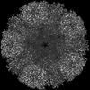
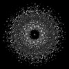
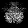
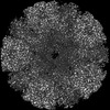
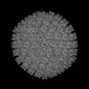
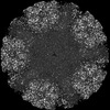
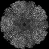
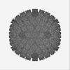
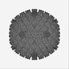
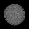
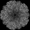
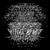
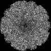
| Title | One CVSC-binding penton vertex in HCMV B-capsid | |||||||||||||||||||||||||||||||||||||||
 Components Components |
| |||||||||||||||||||||||||||||||||||||||
 Keywords Keywords | VIRAL PROTEIN / B-capsid / one CVSC-binding penton vertex / asymmetric reconstruction / VIRUS | |||||||||||||||||||||||||||||||||||||||
| Function / homology |  Function and homology information Function and homology informationT=16 icosahedral viral capsid / viral genome packaging / viral capsid assembly / viral process / viral release from host cell / chromosome organization / viral capsid / host cell nucleus / structural molecule activity / DNA binding Similarity search - Function | |||||||||||||||||||||||||||||||||||||||
| Biological species |   Human betaherpesvirus 5 Human betaherpesvirus 5 | |||||||||||||||||||||||||||||||||||||||
| Method | ELECTRON MICROSCOPY / single particle reconstruction / cryo EM / Resolution: 4.1 Å | |||||||||||||||||||||||||||||||||||||||
 Authors Authors | Li, Z. / Yu, X. | |||||||||||||||||||||||||||||||||||||||
| Funding support |  China, 1items China, 1items
| |||||||||||||||||||||||||||||||||||||||
 Citation Citation | Journal: Nat Commun / Year: 2023 Title: Cryo-electron microscopy structures of capsids and in situ portals of DNA-devoid capsids of human cytomegalovirus. Authors: Zhihai Li / Jingjing Pang / Rongchao Gao / Qingxia Wang / Maoyan Zhang / Xuekui Yu / Abstract: The portal-scaffold complex is believed to nucleate the assembly of herpesvirus procapsids. During capsid maturation, two events occur: scaffold expulsion and DNA incorporation. The portal-scaffold ...The portal-scaffold complex is believed to nucleate the assembly of herpesvirus procapsids. During capsid maturation, two events occur: scaffold expulsion and DNA incorporation. The portal-scaffold interaction and the conformational changes that occur to the portal during the different stages of capsid formation have yet to be elucidated structurally. Here we present high-resolution structures of the A- and B-capsids and in-situ portals of human cytomegalovirus. We show that scaffolds bind to the hydrophobic cavities formed by the dimerization and Johnson-fold domains of the major capsid proteins. We further show that 12 loop-helix-loop fragments-presumably from the scaffold domain-insert into the hydrophobic pocket of the portal crown domain. The portal also undergoes significant changes both positionally and conformationally as it accompanies DNA packaging. These findings unravel the mechanism by which the portal interacts with the scaffold to nucleate capsid assembly and further our understanding of scaffold expulsion and DNA incorporation. | |||||||||||||||||||||||||||||||||||||||
| History |
|
- Structure visualization
| Structure viewer | Molecule: MolmilJmol/JSmol |
|---|
- Downloads & links
-Download
| PDBx/mmCIF format | 8hey.cif.gz | 1.9 MB | Display | PDBx/mmCIF format |
|---|---|---|---|---|
| PDB format | pdb8hey.ent.gz | Display | PDB format | |
| PDBx/mmJSON format | 8hey.json.gz | Tree view | PDBx/mmJSON format | |
| Others |  Other downloads Other downloads |
-Validation report
| Arichive directory | https://data.pdbj.org/pub/pdb/validation_reports/he/8heyftp://data.pdbj.org/pub/pdb/validation_reports/he/8hey | HTTPS FTP |
|---|
-Related structure data
| Related structure data | 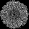 34704MC 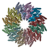 8heuC 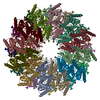 8hevC  8hexC M: map data used to model this data C: citing same article ( |
|---|---|
| Similar structure data |
-Links
 PDBj
PDBj
- Assembly
| Deposited unit | 
|
|---|---|
| 1 | x 5
|
| 2 |
|
| 3 | 
|
| Symmetry | Point symmetry: (Schoenflies symbol: C5 (5 fold cyclic)) |
-Components
-Protein , 2 types, 13 molecules TijQRSaDYZABC
| #1: Protein | Mass: 8495.924 Da / Num. of mol.: 6 / Source method: isolated from a natural source / Source: (natural) Human betaherpesvirus 5 / References: UniProt: A8T7C4#2: Protein | Mass: 154048.906 Da / Num. of mol.: 7 / Source method: isolated from a natural source / Source: (natural) Human betaherpesvirus 5 / References: UniProt: A0A1U8QPG3 |
|---|
-Triplex capsid protein ... , 2 types, 6 molecules hInogm
| #3: Protein | Mass: 34635.750 Da / Num. of mol.: 4 / Source method: isolated from a natural source / Source: (natural) Human betaherpesvirus 5 / References: UniProt: Q6RXF2#4: Protein | Mass: 33071.270 Da / Num. of mol.: 2 / Source method: isolated from a natural source / Source: (natural) Human betaherpesvirus 5 / References: UniProt: Q6RXH2 |
|---|
-Capsid vertex component ... , 2 types, 3 molecules MNO
| #5: Protein | Mass: 68567.211 Da / Num. of mol.: 1 / Source method: isolated from a natural source / Source: (natural) Human betaherpesvirus 5 / References: UniProt: A0A6C0PJD3 |
|---|---|
| #6: Protein | Mass: 71269.570 Da / Num. of mol.: 2 / Source method: isolated from a natural source / Source: (natural) Human betaherpesvirus 5 / References: UniProt: A0A3G6XKK5 |
-Details
| Has protein modification | Y |
|---|
-Experimental details
-Experiment
| Experiment | Method: ELECTRON MICROSCOPY |
|---|---|
| EM experiment | Aggregation state: PARTICLE / 3D reconstruction method: single particle reconstruction |
- Sample preparation
| Component | Name: Human betaherpesvirus 5 / Type: VIRUS / Entity ID: all / Source: NATURAL |
|---|---|
| Source (natural) | Organism: Human betaherpesvirus 5 |
| Details of virus | Empty: NO / Enveloped: YES / Isolate: STRAIN / Type: VIRION |
| Buffer solution | pH: 7.4 |
| Specimen | Embedding applied: NO / Shadowing applied: NO / Staining applied: NO / Vitrification applied: YES |
| Vitrification | Cryogen name: ETHANE |
- Electron microscopy imaging
| Experimental equipment |  Model: Titan Krios / Image courtesy: FEI Company |
|---|---|
| Microscopy | Model: FEI TITAN KRIOS |
| Electron gun | Electron source:  FIELD EMISSION GUN / Accelerating voltage: 300 kV / Illumination mode: FLOOD BEAM FIELD EMISSION GUN / Accelerating voltage: 300 kV / Illumination mode: FLOOD BEAM |
| Electron lens | Mode: BRIGHT FIELD / Nominal defocus max: 2300 nm / Nominal defocus min: 900 nm |
| Image recording | Electron dose: 30 e/Å2 / Film or detector model: GATAN K3 BIOQUANTUM (6k x 4k) |
- Processing
| Software | Name: PHENIX / Version: 1.14_3260: / Classification: refinement |
|---|---|
| EM software | Name: PHENIX / Category: model refinement |
| CTF correction | Type: PHASE FLIPPING AND AMPLITUDE CORRECTION |
| 3D reconstruction | Resolution: 4.1 Å / Resolution method: FSC 0.143 CUT-OFF / Num. of particles: 40903 / Symmetry type: POINT |
