 ムービー
ムービー コントローラー


+ データを開く

- 基本情報
基本情報
| 登録情報 | データベース: EMDB / ID: EMD-3464 | |||||||||
|---|---|---|---|---|---|---|---|---|---|---|
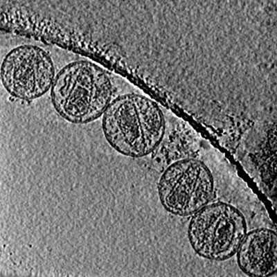
| タイトル | Cryo-electron tomogram containing mature HIV-1 particles | |||||||||
 マップデータ マップデータ | Cryo-electron tomographic reconstruction of wild type mature HIV-1 particles | |||||||||
 試料 試料 |
| |||||||||
| 生物種 |   Human immunodeficiency virus 1 (ヒト免疫不全ウイルス) Human immunodeficiency virus 1 (ヒト免疫不全ウイルス) | |||||||||
| 手法 | 電子線トモグラフィー法 / クライオ電子顕微鏡法 | |||||||||
 データ登録者 データ登録者 | Mattei S / Glass B / Hagen WJH / Kraeusslich H-G / Briggs JAG | |||||||||
 引用 引用 | ジャーナル: Science / 年: 2016 タイトル: The structure and flexibility of conical HIV-1 capsids determined within intact virions. 著者: Simone Mattei / Bärbel Glass / Wim J H Hagen / Hans-Georg Kräusslich / John A G Briggs /  要旨: HIV-1 contains a cone-shaped capsid encasing the viral genome. This capsid is thought to follow fullerene geometry-a curved hexameric lattice of the capsid protein, CA, closed by incorporating 12 CA ...HIV-1 contains a cone-shaped capsid encasing the viral genome. This capsid is thought to follow fullerene geometry-a curved hexameric lattice of the capsid protein, CA, closed by incorporating 12 CA pentamers. Current models for core structure are based on crystallography of hexameric and cross-linked pentameric CA, electron microscopy of tubular CA arrays, and simulations. Here, we report subnanometer-resolution cryo-electron tomography structures of hexameric and pentameric CA within intact HIV-1 particles. Whereas the hexamer structure is compatible with crystallography studies, the pentamer forms using different interfaces. Determining multiple structures revealed how CA flexes to form the variably curved core shell. We show that HIV-1 CA assembles both aberrant and perfect fullerene cones, supporting models in which conical cores assemble de novo after maturation. | |||||||||
| 履歴 |
|
- 構造の表示
構造の表示
| ムービー |
 ムービービューア ムービービューア |
|---|---|
| 構造ビューア | EMマップ: SurfViewMolmilJmol/JSmol |
| 添付画像 |
- ダウンロードとリンク
-EMDBアーカイブ
| マップデータ | emd_3464.map.gz | 632.6 MB | EMDBマップデータ形式 | |
|---|---|---|---|---|
| ヘッダ (付随情報) | emd-3464-v30.xmlemd-3464.xml | 15.6 KB 15.6 KB | 表示 表示 | EMDBヘッダ |
| 画像 |  emd_3464.png emd_3464.png | 143.5 KB | ||
| アーカイブディレクトリ |  http://ftp.pdbj.org/pub/emdb/structures/EMD-3464ftp://ftp.pdbj.org/pub/emdb/structures/EMD-3464 http://ftp.pdbj.org/pub/emdb/structures/EMD-3464ftp://ftp.pdbj.org/pub/emdb/structures/EMD-3464 | HTTPS FTP |
-検証レポート
| 文書・要旨 | emd_3464_validation.pdf.gz | 162.1 KB | 表示 | EMDB検証レポート |
|---|---|---|---|---|
| 文書・詳細版 | emd_3464_full_validation.pdf.gz | 161.2 KB | 表示 | |
| XML形式データ | emd_3464_validation.xml.gz | 5 KB | 表示 | |
| アーカイブディレクトリ | https://ftp.pdbj.org/pub/emdb/validation_reports/EMD-3464ftp://ftp.pdbj.org/pub/emdb/validation_reports/EMD-3464 | HTTPS FTP |
-関連構造データ
| 関連構造データ | 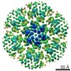 3465C 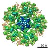 3466C 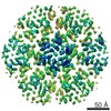 3467C 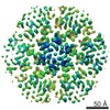 3468C 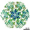 3469C 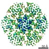 3470C 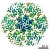 3471C 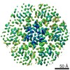 3472C 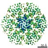 3473C 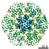 3474C 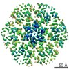 3475C 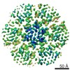 3477C  3478C 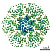 3479C 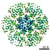 3480C 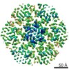 3481C 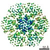 3482C 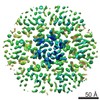 3483C 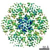 3484C 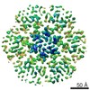 3485C 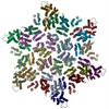 5mcxC 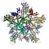 5mcyC  5mczC  5md0C 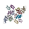 5md1C  5md2C 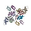 5md3C 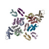 5md4C 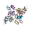 5md5C 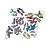 5md6C 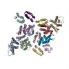 5md7C 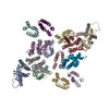 5md8C 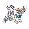 5md9C  5mdaC  5mdbC  5mdcC  5mddC  5mdeC  5mdfC  5mdgC C: 同じ文献を引用 ( |
|---|
-リンク
| EMDBのページ | EMDB (EBI/PDBe) / EMDataResource |
|---|---|
| 「今月の分子」の関連する項目 |

-マップ
| ファイル | ダウンロード / ファイル: emd_3464.map.gz / 形式: CCP4 / 大きさ: 722.7 MB / タイプ: IMAGE STORED AS FLOATING POINT NUMBER (4 BYTES) | ||||||||||||||||||||||||||||||||||||||||||||||||||||||||||||
|---|---|---|---|---|---|---|---|---|---|---|---|---|---|---|---|---|---|---|---|---|---|---|---|---|---|---|---|---|---|---|---|---|---|---|---|---|---|---|---|---|---|---|---|---|---|---|---|---|---|---|---|---|---|---|---|---|---|---|---|---|---|
| 注釈 | Cryo-electron tomographic reconstruction of wild type mature HIV-1 particles | ||||||||||||||||||||||||||||||||||||||||||||||||||||||||||||
| 投影像・断面図 | 画像のコントロール
画像は Spider により作成 これらの図は立方格子座標系で作成されたものです | ||||||||||||||||||||||||||||||||||||||||||||||||||||||||||||
| ボクセルのサイズ | X=Y=Z: 1.78 Å | ||||||||||||||||||||||||||||||||||||||||||||||||||||||||||||
| 密度 |
| ||||||||||||||||||||||||||||||||||||||||||||||||||||||||||||
| 対称性 | 空間群: 1 | ||||||||||||||||||||||||||||||||||||||||||||||||||||||||||||
| 詳細 | EMDB XML:
CCP4マップ ヘッダ情報:
| ||||||||||||||||||||||||||||||||||||||||||||||||||||||||||||
 Z (Sec.)
 Y (Row.)
 X (Col.)


-添付データ
- 試料の構成要素
-全体 : Human immunodeficiency virus 1
| 全体 | 名称: Human immunodeficiency virus 1 (ヒト免疫不全ウイルス) |
|---|---|
| 要素 |
|
-超分子 #1: Human immunodeficiency virus 1
| 超分子 | 名称: Human immunodeficiency virus 1 / タイプ: virus / ID: 1 / 親要素: 0 / 含まれる分子: #1 詳細: HIV-1 particles were produced by infection of MT-4 cells with HIV-1 strain NL43 by coculture. NCBI-ID: 11676 / 生物種: Human immunodeficiency virus 1 / ウイルスタイプ: VIRION / ウイルス・単離状態: STRAIN / ウイルス・エンベロープ: Yes / ウイルス・中空状態: No |
|---|---|
| 宿主 | 生物種:  Homo sapiens (ヒト) Homo sapiens (ヒト) |
-実験情報
-構造解析
| 手法 | クライオ電子顕微鏡法 |
|---|---|
 解析 解析 | 電子線トモグラフィー法 |
| 試料の集合状態 | particle |
-試料調製
| 緩衝液 | pH: 7.4 / 構成要素 - 名称: PBS |
|---|---|
| グリッド | モデル: C-flat / 材質: COPPER / メッシュ: 300 / 支持フィルム - 材質: CARBON / 支持フィルム - トポロジー: HOLEY / 前処理 - タイプ: GLOW DISCHARGE / 前処理 - 雰囲気: AIR |
| 凍結 | 凍結剤: ETHANE / チャンバー内湿度: 100 % / チャンバー内温度: 292 K / 装置: FEI VITROBOT MARK II 詳細: 10nm colloidal gold was added to the sample prior to plunge freezing. |
| 詳細 | Viral particles were purified from cell culture supernatant by iodixanol gradient centrifugation. |
| 切片作成 | その他: NO SECTIONING |
| 位置合わせマーカー | Manufacturer: CMC (Utrecht) / 直径: 10 nm |
- 電子顕微鏡法
| 顕微鏡 | FEI TITAN KRIOS |
|---|---|
| 特殊光学系 | エネルギーフィルター - 名称: GIF Quantum LS エネルギーフィルター - エネルギー下限: 0 eV エネルギーフィルター - エネルギー上限: 20 eV |
| 詳細 | Nanoprobe |
| 撮影 | フィルム・検出器のモデル: GATAN K2 QUANTUM (4k x 4k) 検出モード: SUPER-RESOLUTION / デジタル化 - サイズ - 横: 3710 pixel / デジタル化 - サイズ - 縦: 3838 pixel / 撮影したグリッド数: 1 / 平均電子線量: 2.2 e/Å2 |
| 電子線 | 加速電圧: 300 kV / 電子線源:  FIELD EMISSION GUN FIELD EMISSION GUN |
| 電子光学系 | C2レンズ絞り径: 50.0 µm / 照射モード: FLOOD BEAM / 撮影モード: BRIGHT FIELD / Cs: 2.7 mm / 最大 デフォーカス(公称値): 6.5 µm / 最小 デフォーカス(公称値): 2.0 µm / 倍率(公称値): 81000 |
| 試料ステージ | 試料ホルダーモデル: FEI TITAN KRIOS AUTOGRID HOLDER ホルダー冷却材: NITROGEN |
| 実験機器 |  モデル: Titan Krios / 画像提供: FEI Company |
-画像解析
| 詳細 | Frames were aligned using MotionCorr. Tilts in a tilt series were exposure filtered for cumulative electron dose. Tomograms were reconstructed using IMOD. |
|---|---|
| 最終 再構成 | ソフトウェア - 名称: IMOD / 使用した粒子像数: 45 |
| CTF補正 | ソフトウェア - 名称: IMOD 詳細: CTF correction was performed using the ctfphaseflip program in IMOD prior to backprojection. |
