Movie
Movie Controller

[English] 日本語
 Yorodumi
- EMDB-34529: Un-sharpened map of phycobilisome from Porphyridium purpureum und... -
+ Open data

- Basic information
| Entry | 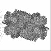 | |||||||||
|---|---|---|---|---|---|---|---|---|---|---|
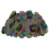
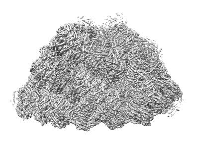
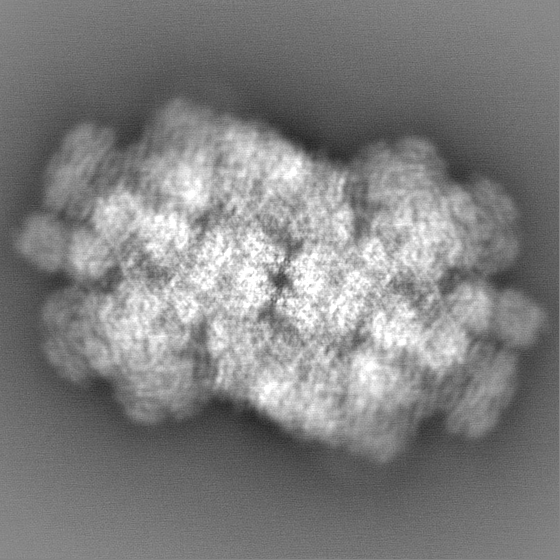
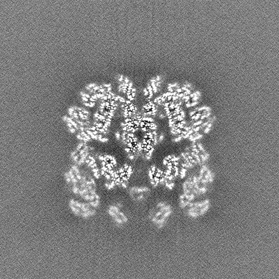
| Title | Un-sharpened map of phycobilisome from Porphyridium purpureum under low light | |||||||||
 Map data Map data | Low Light unsharpen map | |||||||||
 Sample Sample |
| |||||||||
 Keywords Keywords | phycobilisome / Complex / PHOTOSYNTHESIS | |||||||||
| Biological species |  Porphyridium purpureum (eukaryote) Porphyridium purpureum (eukaryote) | |||||||||
| Method | single particle reconstruction / cryo EM / Resolution: 3.74 Å | |||||||||
 Authors Authors | Sui SF / Ma JF | |||||||||
| Funding support |  China, 1 items China, 1 items
| |||||||||
 Citation Citation | Journal: Nature / Year: 2020 Title: Structural basis of energy transfer in Porphyridium purpureum phycobilisome. Authors: Jianfei Ma / Xin You / Shan Sun / Xiaoxiao Wang / Song Qin / Sen-Fang Sui / Abstract: Photosynthetic organisms have developed various light-harvesting systems to adapt to their environments. Phycobilisomes are large light-harvesting protein complexes found in cyanobacteria and red ...Photosynthetic organisms have developed various light-harvesting systems to adapt to their environments. Phycobilisomes are large light-harvesting protein complexes found in cyanobacteria and red algae, although how the energies of the chromophores within these complexes are modulated by their environment is unclear. Here we report the cryo-electron microscopy structure of a 14.7-megadalton phycobilisome with a hemiellipsoidal shape from the red alga Porphyridium purpureum. Within this complex we determine the structures of 706 protein subunits, including 528 phycoerythrin, 72 phycocyanin, 46 allophycocyanin and 60 linker proteins. In addition, 1,598 chromophores are resolved comprising 1,430 phycoerythrobilin, 48 phycourobilin and 120 phycocyanobilin molecules. The markedly improved resolution of our structure compared with that of the phycobilisome of Griffithsia pacifica enabled us to build an accurate atomic model of the P. purpureum phycobilisome system. The model reveals how the linker proteins affect the microenvironment of the chromophores, and suggests that interactions of the aromatic amino acids of the linker proteins with the chromophores may be a key factor in fine-tuning the energy states of the chromophores to ensure the efficient unidirectional transfer of energy. | |||||||||
| History |
|
- Structure visualization
| Supplemental images |
|---|
- Downloads & links
-EMDB archive
| Map data | emd_34529.map.gz | 606.8 MB |  EMDB map data format EMDB map data format | |
|---|---|---|---|---|
| Header (meta data) | emd-34529-v30.xmlemd-34529.xml | 18.5 KB 18.5 KB | Display Display | EMDB header |
| Images |  emd_34529.png emd_34529.png | 116.4 KB | ||
| Filedesc metadata | emd-34529.cif.gz | 4.2 KB | ||
| Others | emd_34529_half_map_1.map.gzemd_34529_half_map_2.map.gz | 614.2 MB 614.2 MB | ||
| Archive directory |  http://ftp.pdbj.org/pub/emdb/structures/EMD-34529ftp://ftp.pdbj.org/pub/emdb/structures/EMD-34529 http://ftp.pdbj.org/pub/emdb/structures/EMD-34529ftp://ftp.pdbj.org/pub/emdb/structures/EMD-34529 | HTTPS FTP |
-Related structure data
| Related structure data | 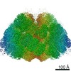 9976C 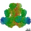 9977C 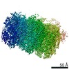 9978C 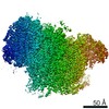 9979C 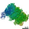 9980C 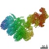 9981C 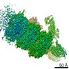 9982C 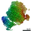 9983C 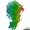 9984C 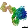 9985C 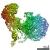 9986C 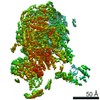 9987C 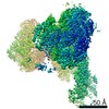 9988C 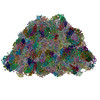 6kgxC C: citing same article ( |
|---|
-Links
| EMDB pages | EMDB (EBI/PDBe) / EMDataResource |
|---|
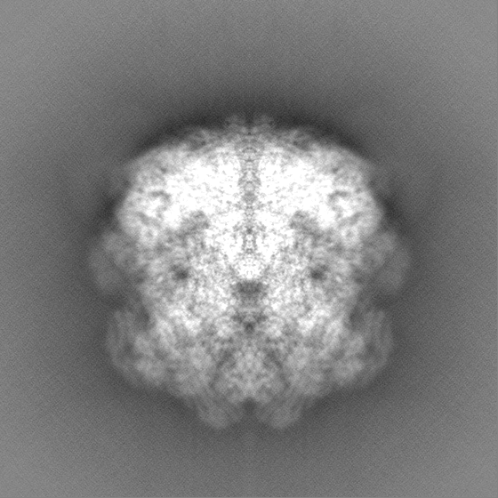
-Map
| File | Download / File: emd_34529.map.gz / Format: CCP4 / Size: 669.9 MB / Type: IMAGE STORED AS FLOATING POINT NUMBER (4 BYTES) | ||||||||||||||||||||||||||||||||||||
|---|---|---|---|---|---|---|---|---|---|---|---|---|---|---|---|---|---|---|---|---|---|---|---|---|---|---|---|---|---|---|---|---|---|---|---|---|---|
| Annotation | Low Light unsharpen map | ||||||||||||||||||||||||||||||||||||
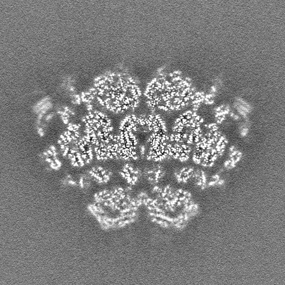
| Projections & slices | Image control
Images are generated by Spider. | ||||||||||||||||||||||||||||||||||||
| Voxel size | X=Y=Z: 1.091 Å | ||||||||||||||||||||||||||||||||||||
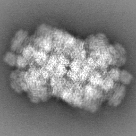
| Density |
| ||||||||||||||||||||||||||||||||||||
| Symmetry | Space group: 1 | ||||||||||||||||||||||||||||||||||||
| Details | EMDB XML:
|
 Z (Sec.)
 Y (Row.)
 X (Col.)


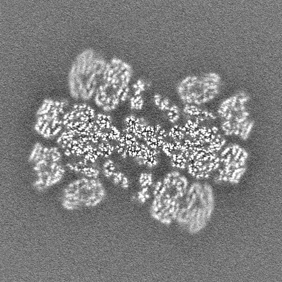
-Supplemental data
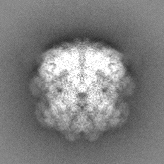
-Half map: Low Light unsharpen map half2
| File | emd_34529_half_map_1.map | ||||||||||||
|---|---|---|---|---|---|---|---|---|---|---|---|---|---|
| Annotation | Low Light unsharpen map_half2 | ||||||||||||
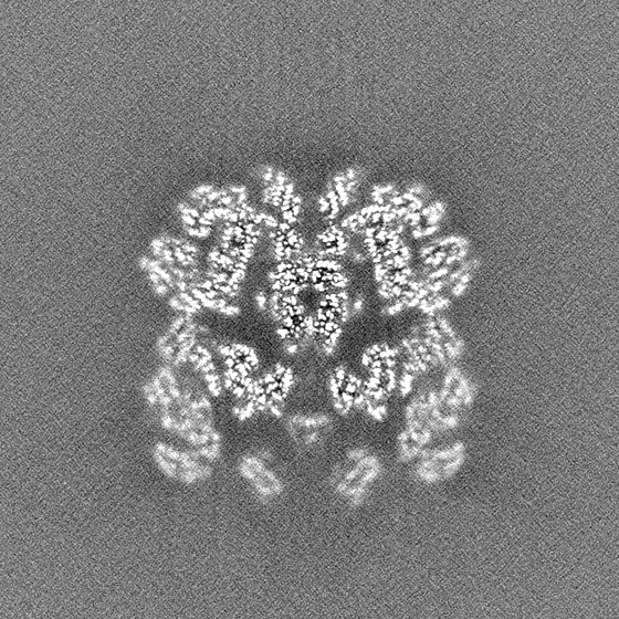
| Projections & Slices |
| ||||||||||||
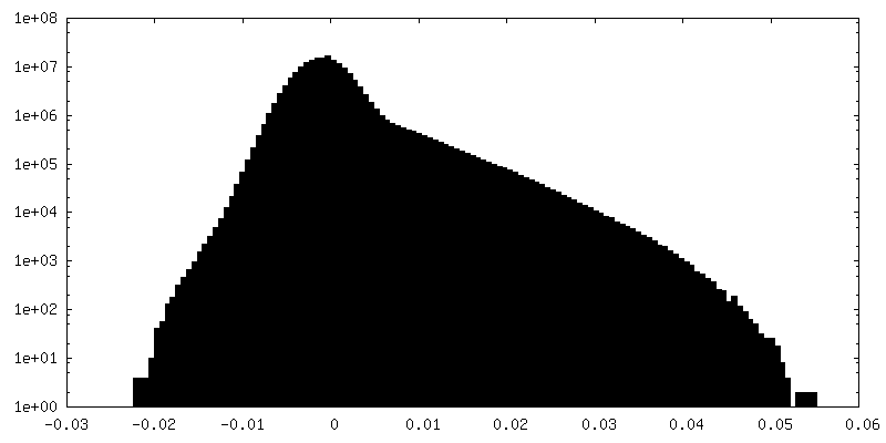
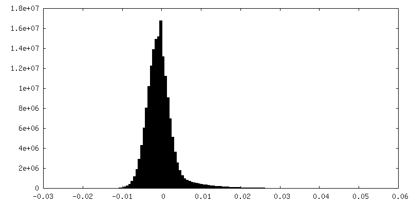
| Density Histograms |

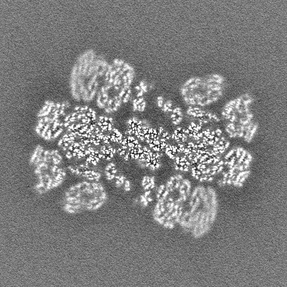

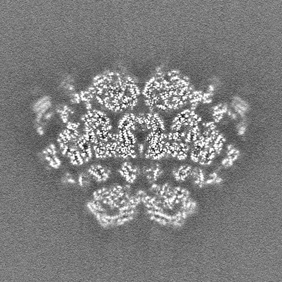
-Half map: Low Light unsharpen map half1
| File | emd_34529_half_map_2.map | ||||||||||||
|---|---|---|---|---|---|---|---|---|---|---|---|---|---|
| Annotation | Low Light unsharpen map_half1 | ||||||||||||
| Projections & Slices |
| ||||||||||||
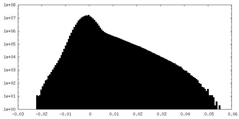
| Density Histograms |

- Sample components
-Entire : Complex of phycobilisome from red alga P.purpureum.
| Entire | Name: Complex of phycobilisome from red alga P.purpureum. |
|---|---|
| Components |
|
-Supramolecule #1: Complex of phycobilisome from red alga P.purpureum.
| Supramolecule | Name: Complex of phycobilisome from red alga P.purpureum. / type: complex / ID: 1 / Parent: 0 / Macromolecule list: #1-#25 |
|---|---|
| Source (natural) | Organism: Porphyridium purpureum (eukaryote) |
-Experimental details
-Structure determination
| Method | cryo EM |
|---|---|
 Processing Processing | single particle reconstruction |
| Aggregation state | particle |
-Sample preparation
| Concentration | 1.5 mg/mL | |||||||||
|---|---|---|---|---|---|---|---|---|---|---|
| Buffer | pH: 7 Component:
| |||||||||
| Sugar embedding | Material: ice | |||||||||
| Grid | Model: Quantifoil R2/2 / Support film - Material: CARBON / Support film - topology: CONTINUOUS / Support film - Film thickness: 5 | |||||||||
| Vitrification | Cryogen name: ETHANE / Chamber humidity: 100 % / Chamber temperature: 298 K |
- Electron microscopy
| Microscope | FEI TITAN KRIOS |
|---|---|
| Specialist optics | Energy filter - Name: GIF Quantum LS / Energy filter - Slit width: 20 eV |
| Image recording | Film or detector model: GATAN K2 SUMMIT (4k x 4k) / Detector mode: SUPER-RESOLUTION / Digitization - Frames/image: 1-32 / Number real images: 16218 / Average exposure time: 5.6 sec. / Average electron dose: 48.0 e/Å2 |
| Electron beam | Acceleration voltage: 300 kV / Electron source:  FIELD EMISSION GUN FIELD EMISSION GUN |
| Electron optics | C2 aperture diameter: 70.0 µm / Illumination mode: OTHER / Imaging mode: BRIGHT FIELD / Cs: 0.001 mm / Nominal defocus max: 2.2 µm / Nominal defocus min: 1.2 µm / Nominal magnification: 105000 |
| Sample stage | Specimen holder model: FEI TITAN KRIOS AUTOGRID HOLDER / Cooling holder cryogen: NITROGEN |
| Experimental equipment |  Model: Titan Krios / Image courtesy: FEI Company |