 Movie
Movie Controller

[English] 日本語
 Yorodumi
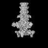
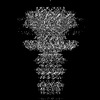
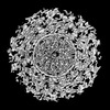
- EMDB-34252: Cryo-EM model of the marine siphophage vB_DshS-R4C stopper-termin... -
+ Open data

- Basic information
| Entry |  | |||||||||
|---|---|---|---|---|---|---|---|---|---|---|
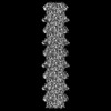
| Title | Cryo-EM model of the marine siphophage vB_DshS-R4C stopper-terminator complex | |||||||||
 Map data Map data | ||||||||||
 Sample Sample |
| |||||||||
 Keywords Keywords | Marine bacteriophage / Cryo-EM / Siphophage / Stopper protein / Terminator protein / Head-to-tail interface / VIRUS | |||||||||
| Function / homology | Bacteriophage tail attachment protein FII / : / Phage Head-Tail Attachment protein / virion assembly / Head-to-tail joining protein / Minor tail protein / Major tail protein Function and homology information Function and homology information | |||||||||
| Biological species |  Dinoroseobacter phage vB_DshS-R4C (virus) Dinoroseobacter phage vB_DshS-R4C (virus) | |||||||||
| Method | single particle reconstruction / cryo EM / Resolution: 6.6 Å | |||||||||
 Authors Authors | Huang Y / Sun H / Wei S / Zheng Q / Li S / Zhang R / Xia N | |||||||||
| Funding support | 1 items
| |||||||||
 Citation Citation | Journal: Nat Commun / Year: 2023 Title: Structure and proposed DNA delivery mechanism of a marine roseophage. Authors: Yang Huang / Hui Sun / Shuzhen Wei / Lanlan Cai / Liqin Liu / Yanan Jiang / Jiabao Xin / Zhenqin Chen / Yuqiong Que / Zhibo Kong / Tingting Li / Hai Yu / Jun Zhang / Ying Gu / Qingbing Zheng ...Authors: Yang Huang / Hui Sun / Shuzhen Wei / Lanlan Cai / Liqin Liu / Yanan Jiang / Jiabao Xin / Zhenqin Chen / Yuqiong Que / Zhibo Kong / Tingting Li / Hai Yu / Jun Zhang / Ying Gu / Qingbing Zheng / Shaowei Li / Rui Zhang / Ningshao Xia /  Abstract: Tailed bacteriophages (order, Caudovirales) account for the majority of all phages. However, the long flexible tail of siphophages hinders comprehensive investigation of the mechanism of viral gene ...Tailed bacteriophages (order, Caudovirales) account for the majority of all phages. However, the long flexible tail of siphophages hinders comprehensive investigation of the mechanism of viral gene delivery. Here, we report the atomic capsid and in-situ structures of the tail machine of the marine siphophage, vB_DshS-R4C (R4C), which infects Roseobacter. The R4C virion, comprising 12 distinct structural protein components, has a unique five-fold vertex of the icosahedral capsid that allows genome delivery. The specific position and interaction pattern of the tail tube proteins determine the atypical long rigid tail of R4C, and further provide negative charge distribution within the tail tube. A ratchet mechanism assists in DNA transmission, which is initiated by an absorption device that structurally resembles the phage-like particle, RcGTA. Overall, these results provide in-depth knowledge into the intact structure and underlining DNA delivery mechanism for the ecologically important siphophages. | |||||||||
| History |
|
- Structure visualization
| Supplemental images |
|---|

- Downloads & links
-EMDB archive
| Map data | emd_34252.map.gz | 49.1 MB | EMDB map data format | |
|---|---|---|---|---|
| Header (meta data) | emd-34252-v30.xmlemd-34252.xml | 16 KB 16 KB | Display Display | EMDB header |
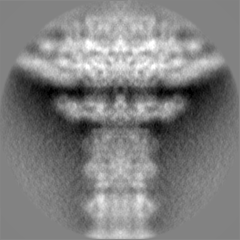
| Images | 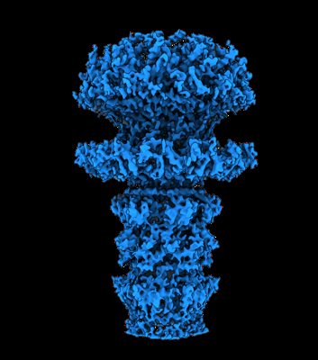 emd_34252.png emd_34252.png | 109.4 KB | ||
| Filedesc metadata | emd-34252.cif.gz | 5.4 KB | ||
| Others | emd_34252_half_map_1.map.gzemd_34252_half_map_2.map.gz | 39.5 MB 39.5 MB | ||
| Archive directory |  http://ftp.pdbj.org/pub/emdb/structures/EMD-34252ftp://ftp.pdbj.org/pub/emdb/structures/EMD-34252 http://ftp.pdbj.org/pub/emdb/structures/EMD-34252ftp://ftp.pdbj.org/pub/emdb/structures/EMD-34252 | HTTPS FTP |
-Validation report
| Summary document | emd_34252_validation.pdf.gz | 1.1 MB | Display | EMDB validaton report |
|---|---|---|---|---|
| Full document | emd_34252_full_validation.pdf.gz | 1.1 MB | Display | |
| Data in XML | emd_34252_validation.xml.gz | 11.8 KB | Display | |
| Data in CIF | emd_34252_validation.cif.gz | 13.8 KB | Display | |
| Arichive directory | https://ftp.pdbj.org/pub/emdb/validation_reports/EMD-34252ftp://ftp.pdbj.org/pub/emdb/validation_reports/EMD-34252 | HTTPS FTP |
-Related structure data
| Related structure data |  8gtfMC  8gtaC 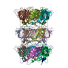 8gtbC  8gtcC 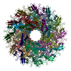 8gtdC M: atomic model generated by this map C: citing same article ( |
|---|---|
| Similar structure data |
-Links
| EMDB pages | EMDB (EBI/PDBe) / EMDataResource |
|---|
-Map
| File | Download / File: emd_34252.map.gz / Format: CCP4 / Size: 52.7 MB / Type: IMAGE STORED AS FLOATING POINT NUMBER (4 BYTES) | ||||||||||||||||||||||||||||||||||||
|---|---|---|---|---|---|---|---|---|---|---|---|---|---|---|---|---|---|---|---|---|---|---|---|---|---|---|---|---|---|---|---|---|---|---|---|---|---|
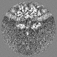
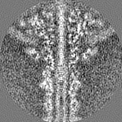
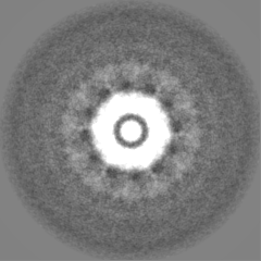
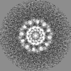
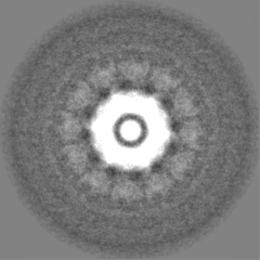
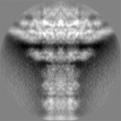
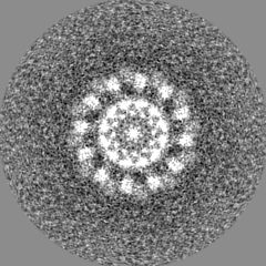
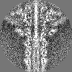
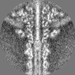
| Projections & slices | Image control
Images are generated by Spider. | ||||||||||||||||||||||||||||||||||||
| Voxel size | X=Y=Z: 1.128 Å | ||||||||||||||||||||||||||||||||||||
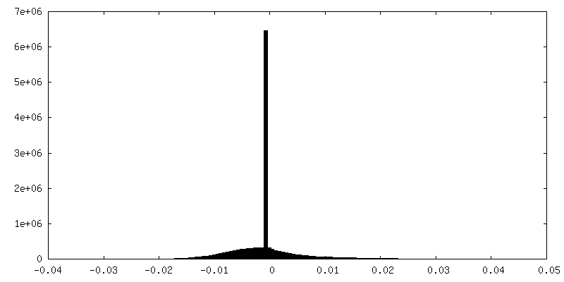
| Density |
| ||||||||||||||||||||||||||||||||||||
| Symmetry | Space group: 1 | ||||||||||||||||||||||||||||||||||||
| Details | EMDB XML:
|
 Z (Sec.)
 Y (Row.)
 X (Col.)


-Supplemental data
-Half map: #2
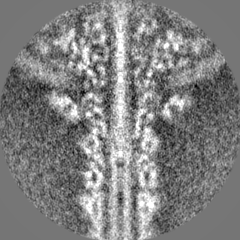
| File | emd_34252_half_map_1.map | ||||||||||||
|---|---|---|---|---|---|---|---|---|---|---|---|---|---|
| Projections & Slices |
| ||||||||||||
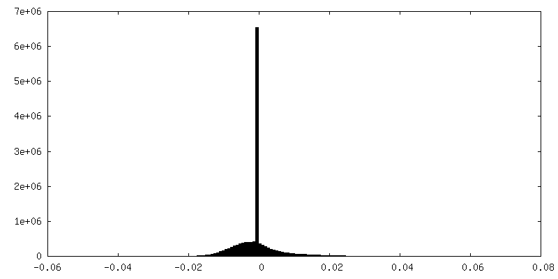
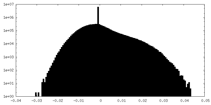
| Density Histograms |

-Half map: #1
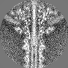
| File | emd_34252_half_map_2.map | ||||||||||||
|---|---|---|---|---|---|---|---|---|---|---|---|---|---|
| Projections & Slices |
| ||||||||||||
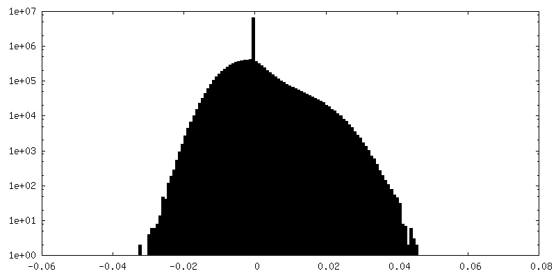
| Density Histograms |

- Sample components
-Entire : Dinoroseobacter phage vB_DshS-R4C
| Entire | Name: Dinoroseobacter phage vB_DshS-R4C (virus) |
|---|---|
| Components |
|
-Supramolecule #1: Dinoroseobacter phage vB_DshS-R4C
| Supramolecule | Name: Dinoroseobacter phage vB_DshS-R4C / type: virus / ID: 1 / Parent: 0 / Macromolecule list: all / NCBI-ID: 2590919 / Sci species name: Dinoroseobacter phage vB_DshS-R4C / Virus type: VIRION / Virus isolate: SPECIES / Virus enveloped: No / Virus empty: No |
|---|---|
| Host (natural) | Organism:  Dinoroseobacter shibae DFL 12 = DSM 16493 (bacteria) Dinoroseobacter shibae DFL 12 = DSM 16493 (bacteria) |
-Macromolecule #1: Head-to-tail joining protein
| Macromolecule | Name: Head-to-tail joining protein / type: protein_or_peptide / ID: 1 / Number of copies: 6 / Enantiomer: LEVO |
|---|---|
| Source (natural) | Organism: Dinoroseobacter phage vB_DshS-R4C (virus) |
| Molecular weight | Theoretical: 10.946397 KDa |
| Sequence | String: MIESLADWSI FTDPDVFGEP VTWTTPPLPD PVPAIFTDAS EDRPATLGPG VLTIAPTLTL GAAQLPFSPA RNHRCTVRGI TYRVAEVQP DGSGGLRLLL ERV UniProtKB: Head-to-tail joining protein |
-Macromolecule #2: Major tail protein
| Macromolecule | Name: Major tail protein / type: protein_or_peptide / ID: 2 / Number of copies: 6 / Enantiomer: LEVO |
|---|---|
| Source (natural) | Organism: Dinoroseobacter phage vB_DshS-R4C (virus) |
| Molecular weight | Theoretical: 13.574816 KDa |
| Sequence | String: MLKGKDGVVK NASTGDSIGH LQSWALDTQR DEVSGWGMGD DAERAFTTVG RASGNFEVYL DPADPSDDLE PGDLVDLELY PGGESTGSG YRSVAGALIL STAESASKDG IPMLTVNWRT SGALPQKATV S UniProtKB: Major tail protein |
-Macromolecule #3: Terminator protein
| Macromolecule | Name: Terminator protein / type: protein_or_peptide / ID: 3 / Number of copies: 6 / Enantiomer: LEVO |
|---|---|
| Source (natural) | Organism: Dinoroseobacter phage vB_DshS-R4C (virus) |
| Molecular weight | Theoretical: 14.866825 KDa |
| Sequence | String: MSEAIIAAAR GRLISPPFSD ATGDVYRTPE AALPAIIVEL DYTDAERISM GGGFIASAEL RVEILAKRDD WSLLTPTPAN TAEGMARLA ALVRTAILAP PSDLSGLAWS IAPAGYEFET ERGETPLARA TQSFALQILQ P UniProtKB: Minor tail protein |
-Experimental details
-Structure determination
| Method | cryo EM |
|---|---|
 Processing Processing | single particle reconstruction |
| Aggregation state | particle |
-Sample preparation
| Buffer | pH: 7.6 |
|---|---|
| Vitrification | Cryogen name: ETHANE |
- Electron microscopy
| Microscope | FEI TECNAI F30 |
|---|---|
| Image recording | Film or detector model: FEI FALCON III (4k x 4k) / Average electron dose: 30.0 e/Å2 |
| Electron beam | Acceleration voltage: 300 kV / Electron source:  FIELD EMISSION GUN FIELD EMISSION GUN |
| Electron optics | Illumination mode: FLOOD BEAM / Imaging mode: BRIGHT FIELD / Nominal defocus max: 2.2 µm / Nominal defocus min: 1.0 µm |
| Experimental equipment |  Model: Tecnai F30 / Image courtesy: FEI Company |
-Image processing
| Startup model | Type of model: OTHER / Details: trRosetta server |
|---|---|
| Final reconstruction | Applied symmetry - Point group: C6 (6 fold cyclic) / Resolution.type: BY AUTHOR / Resolution: 6.6 Å / Resolution method: FSC 0.143 CUT-OFF / Number images used: 5844 |
| Initial angle assignment | Type: MAXIMUM LIKELIHOOD |
| Final angle assignment | Type: MAXIMUM LIKELIHOOD |
